不同分娩方式對產后早期盆底肌電值及肌力的影響
2019-10-09 12:11:12聶偉劉學云張琳琳黑國真
中國醫藥導報 2019年20期
關鍵詞:剖宮產術
聶偉 劉學云 張琳琳 黑國真
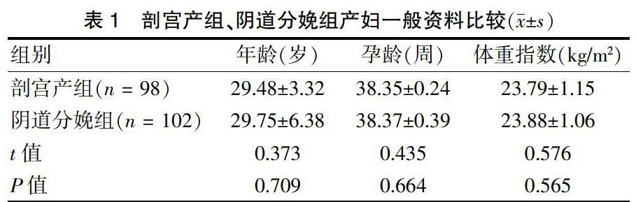
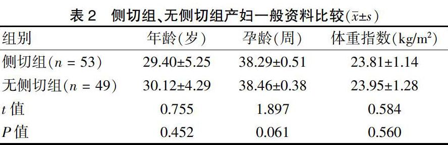
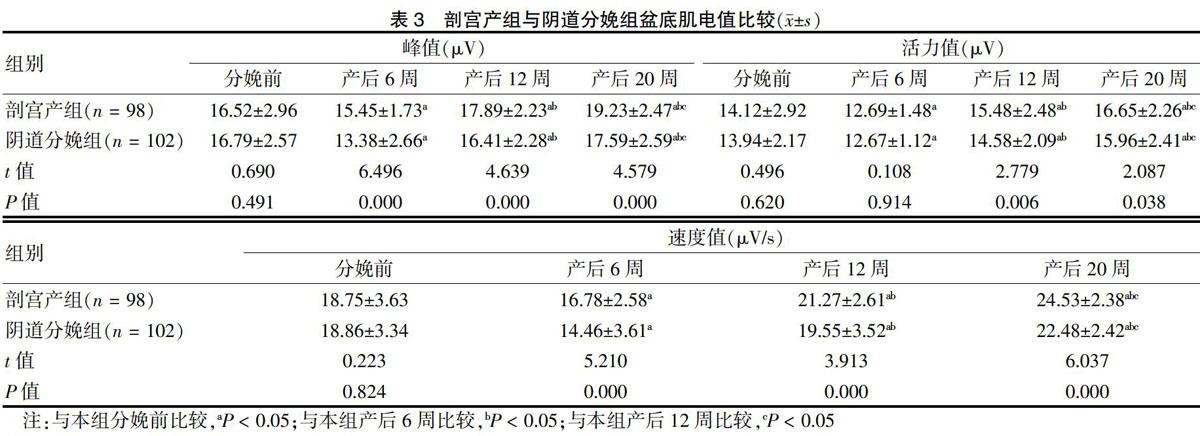
[摘要] 目的 探討不同分娩方式對產后早期盆底肌電值及肌力的影響。 方法 選取2015年6月~2018年10月于山東省聊城市東昌府區婦幼保健院產檢并分娩的200例產婦為研究對象,按照分娩方式的不同將其分為剖宮產組(n = 98)和陰道分娩組(n = 102),陰道分娩組根據有無側切分為側切組(n = 53)和無側切組(n = 49)。分別于分娩前、產后6周、產后12周、產后20周,比較剖宮產組與陰道分娩組、側切組和無側切組組間的盆底肌電值和肌力下降情況。 結果 產后6、12、20周,兩組盆底肌電位峰值、活力值、速度值均呈先降低后升高趨勢(P < 0.05);產后6周,剖宮產組除盆底肌電位活力值與陰道分娩組比較差異無統計學意義(P > 0.05)外,其余各時間點的盆底肌電位峰值、活力值、速度值均高于陰道分娩組(P < 0.05)。分娩前及產后6、12、20周,側切組與無側切組盆底肌電位峰值、活力值、速度值等比較,差異無統計學意義(P > 0.05),產后6周兩組電位峰值、活力值、速度值均低于分娩前(P < 0.05)。分娩前及產后6、12、20周,剖宮產組、陰道分娩組以及側切組、無側切組Ⅰ類肌、Ⅱ類肌肌力下降患者占比比較,差異均無統計學意義(P > 0.05)。 結論 剖宮產、陰道分娩對盆底肌力下降的影響區別不大,且陰道分娩中側切不能減輕對產后盆底肌力的影響。剖宮產對產后早期盆底肌電值的影響較陰道分娩小。
[關鍵詞] 剖宮產術;自然分娩;會陰切開術;產后;盆肌電值;肌力
[中圖分類號] R714.254? ? ? ? ? [文獻標識碼] A? ? ? ? ? [文章編號] 1673-7210(2019)07(b)-0095-05
Effect of different delivery modes on pelvic floor electromyography and muscle strength in early postpartum period
NIE Wei1? ?LIU Xueyun2? ?ZHANG Linlin3? ?HEI Guozhen3
1.Research Department, Shandong Academy of Medical Sciences, Shandong Province, Ji′nan? ?250000, China; 2.Department of Obstetrics, Shandong Provincial Qianfoshan Hospital, Shandong Province, Ji′nan? ?250000, China; 3.Department of Obstetrics, Liaocheng Dongchangfu District Maternal and Child Health Hospital, Shandong Province, Liaocheng? ?252000, China
[Abstract] Objective To investigate the effect of different delivery methods on pelvic floor electromyography and muscle strength in early postpartum period. Methods Total of 200 parturients who were examined and delivered in Liaocheng Dongchangfu District Maternal and Child Health Hospital of Shandong Province from June 2015 to October 2018 were selected as the research objects. They were divided into cesarean section group (n = 98) and vaginal delivery group (n = 102), vaginal delivery group was divided into lateral section group (n = 53) and non-lateral section group (n = 49) according to the different modes of delivery. The pelvic floor myoelectricity and muscle strength were compared between cesarean section group and vaginal delivery group, lateral incision group and the non-lateral incision group before delivery, 6, 12 and 20 weeks postpartum. Results The peak value, activity value and velocity value of pelvic floor potential in cesarean section group and vaginal delivery group decreased first and then increased at 6,12 and 20 weeks postpartum (P < 0.05). There was no significant difference in pelvic floor potential activity between cesarean section group and vaginal delivery group at 6 weeks postpartum (P > 0.05). The peak value of pelvic floor muscle potential, pelvic floor muscle activity and pelvic floor velocity at other time points were higher than those in vaginal delivery group (P < 0.05). There was no statistical difference in peak pelvic floor potential, pelvic floor activity and pelvic floor velocity between the two groups (P > 0.05), and the peak pelvic floor potential, pelvic floor activity and pelvic floor velocity of the two groups was lower 6 weeks postpartum than those before delivery (P < 0.05). There was no statistical difference in the decrease rate of muscular strength of type Ⅰ and Ⅱ muscles between cesarean section group, vaginal delivery group and lateral section group and non-lateral section group (P > 0.05). Conclusion The influence of cesarean section and vaginal delivery on the decrease of pelvic floor muscle strength is not different, and lateral incision during vaginal delivery can not reduce the influence of pelvic floor muscle strength after delivery. The effect of cesarean section on pelvic floor EMG in early postpartum period is slight.
猜你喜歡
中國當代醫藥(2016年30期)2017-01-07 00:57:09
中國實用醫藥(2016年30期)2016-12-28 09:08:45
中國實用醫藥(2016年18期)2016-08-03 09:23:02
中國實用醫藥(2016年16期)2016-07-26 19:49:02
中國實用醫藥(2016年17期)2016-07-26 13:39:19
中國實用醫藥(2016年13期)2016-07-05 04:03:26
中國實用醫藥(2016年11期)2016-05-04 16:08:28
中國實用醫藥(2016年10期)2016-05-04 11:15:59
中國科技博覽(2016年5期)2016-04-23 20:09:54
中國實用醫藥(2016年8期)2016-03-30 23:48:51