Ⅵ型膠原在正常軟骨和骨關節炎軟骨中的空間分布
2015-08-29 01:20:09王恒沙常童潔高瑩瑩張里程張立海唐佩福
解放軍醫學院學報 2015年7期
關鍵詞:骨關節炎
李 曌,李 鵬,王恒沙,常童潔,高瑩瑩,陳 超,張里程,張立海,張 毅,唐佩福
1解放軍總醫院,北京 100853;2軍事醫學科學院 基礎醫學研究所細胞生物學研究室,北京 100850;3北京雪邦科技有限公司,北京 100039;4清華大學蛋白質設施細胞影像平臺,北京 100084
Ⅵ型膠原在正常軟骨和骨關節炎軟骨中的空間分布
李 曌1,2,李 鵬1,2,王恒沙3,常童潔4,高瑩瑩4,陳 超1,張里程1,張立海1,張 毅2,唐佩福1
1解放軍總醫院,北京 100853;2軍事醫學科學院 基礎醫學研究所細胞生物學研究室,北京 100850;3北京雪邦科技有限公司,北京 100039;4清華大學蛋白質設施細胞影像平臺,北京 100084
目的 探究骨關節炎軟骨中Ⅵ型膠原空間分布的變化規律。方法 應用Hartley豚鼠自發性骨關節炎模型(模型組)和健康豚鼠(對照組),取膝關節軟骨做冷凍切片,進行Ⅵ型膠原免疫熒光染色,利用Delta-Vision Elite成像系統和Imaris軟件進行斷層掃描、3D重建和定量分析,比較兩組軟骨細胞體積、Ⅵ型膠原厚度與體積的差異。結果 正常軟骨中,薄層Ⅵ型膠原層均勻包裹軟骨細胞,各層細胞外Ⅵ型膠原層厚度無統計學差異(P>0.05),體積隨細胞深度增加而增加(P<0.01);骨關節炎軟骨中,Ⅵ型膠原體積減小(P<0.01)且空間分布不規則,移行層呈蜂窩狀或出現空洞,放射層中呈點、片狀彌散分布,且在細胞周基質外出現散在的Ⅵ型膠原。結論 在骨關節炎軟骨中,Ⅵ型膠原體積減少,不完全包裹軟骨細胞,出現蜂窩、空洞和彌散分布的現象,這種改變可能是軟骨細胞變性的誘因之一。
Ⅵ型膠原;軟骨細胞;骨關節炎;豚鼠自發性骨關節炎模型
網絡出版時間:2015-04-21 09:26 網絡出版地址:http://www.cnki.net/kcms/detail/11.3275.R.20150421.0926.003.html
軟骨細胞維持著關節軟骨的新陳代謝和正常生理功能[1],調控軟骨細胞的物理、化學微環境和維持正常軟骨細胞表型的最重要結構是軟骨細胞周基質(pericellular matrix,PCM)[2],Ⅵ型膠原蛋白(type Ⅵ collagen)是PCM的一種主要成分[3],其連接于軟骨細胞和Ⅱ型膠原網之間,增加了PCM的彈性模量,且與PCM中多種蛋白成分(如纖連蛋白、纖調蛋白等)結合[4],因此被認為是調節軟骨細胞物理、化學微環境的重要物質[5-6]。
軟骨細胞變性是骨關節炎(osteoarthritis,OA)病理演變的關鍵[1],軟骨細胞物理、化學微環境的改變在其中起到重要作用。Pullig等[7]報道了在OA軟骨中Ⅵ型膠原蛋白含量有明顯改變,因此它可能與軟骨細胞退變有關。由于關節軟骨的空間結構對其生物力學性能有很大影響,因此Ⅵ型膠原蛋白空間分布的變化可能在OA發病過程中有重要作用,但目前并無相關文獻報道。因此,本研究利用豚鼠自發性OA模型與健康豚鼠對比[8],通過3D圖像分析OA軟骨中Ⅵ型膠原的空間分布特征,為深入研究OA的病理機制提供基礎。
材料和方法
1 實驗動物 雄性Hartley豚鼠1月齡5只,體質量300 ~ 350 (332.00±18.91) g;12月齡5只,體質量1 000 ~ 1 300 (1 172.00±86.36) g。購自北京維通利華實驗動物中心(許可證號:SCXK(京)2012-0001)。
2 主要試劑與儀器 Frozen Section Compound(Leica);蘇木素-伊紅染色試劑(江蘇碧云天);Anti-CollagenⅥantibody(abcam);FITC-IgG(Santa Cruz);DeltaVision-System(GE),搭載平場復消色差空氣鏡(40×,數值孔徑0.95)、平場復消色差油鏡(60×,數值孔徑1.35)以及軟件SoftWoRx Suite 2.0;Imaris 8.0(Bitplane)。
3 軟骨標本的制備 1月齡豚鼠為健康對照組,12月齡為自發性OA組[9];處死豚鼠后解剖雙下肢,于內側脛骨平臺處,用手術刀片刮取約0.3 cm× 0.5 cm的軟骨片數片,PBS溶液沖洗后于濾紙上吸干表面水分并展平,冷凍包埋劑包埋后-70℃冷凍待用。
4 Ⅵ型膠原免疫熒光染色(immunofluorescence staining,IF) 垂直于軟骨表面切取40 μm冷凍切片[6],3%過氧化氫溶液浸泡60 min,室溫下10%山羊血清封閉20 min,4℃Anti-CollagenⅥ antibody(1∶50)孵育過夜,PBS漂洗3次,室溫下FITC二抗(1∶100)避光孵育30min,PBS漂洗3次后封片待用。以PBS代替一抗作為陰性對照,其他染色方法同上。
5 3D圖像采集及處理 在Delta-Vision成像系統[10]觀察IF切片Ⅵ型膠原的分布;40×鏡下選取典型細胞和區域,60×下(分辨率2 048×2 048,層掃厚度0.2μm)進行細節拍攝,7次去卷積處理后,圖像輸入Imaris8.0[11],Slice模式(2D)下選取典型細胞(每組每帶各10個),測量熒光區域的平均厚度;Surpass模式(3D)下篩選出熒光包裹完整的細胞(每組每帶各10個),Surface模塊下對熒光區域及熒光包裹區域進行表面重建。測量重建后的熒光區域體積(Ⅵ型膠原體積,Vp)、包裹的空腔體積(軟骨細胞體積,Vc)。
6 統計學處理 實驗數據采用SPSS19.0統計軟件進行分析,數據描述為±s,兩組均數比較采用成組設計的t檢驗,多組均數比較采用方差分析,P<0.05為差異有統計學意義。
結 果
1 正常軟骨中Ⅵ型膠原的分布 正常軟骨中Ⅵ型膠原均勻包裹在軟骨細胞表面(圖1A)。表面層Ⅵ型膠原(圖1B)呈盤狀,長軸與細胞表面平行;移行層Ⅵ型膠原(圖1C)近似球體;放射層Ⅵ型膠原包裹的細胞(圖1D)大多成對、成組存在,呈橢球體,長軸方向不定。各層細胞表面Ⅵ型膠原厚度:表面層(1.09±0.16)μm,移行層(1.16±0.26)μm,放射層(1.02±0.12)μm,各層厚度差異無統計學意義(P=0.30)。軟骨細胞(Vc)及其周圍Ⅵ型膠原體積(Vp)測量結果(圖2A):表面層Vc(154.90± 41.90)μm3,Vp(293.20±55.48)μm3;移行層Vc(709.50±156.20)μm3,Vp(1 033.60±152.99)μm3;放射層Vc(992.60±111.08)μm3,Vp(1 193.00± 277.73)μm3。從表面層到放射層Ⅵ型膠原所代表的PCM體積隨軟骨細胞體積的增加而增加(P<0.05),但移行層和放射層Ⅵ型膠原體積差異無統計學意義(P>0.05)。
2 OA軟骨中Ⅵ型膠原的分布 OA軟骨中的細胞形態和Ⅵ型膠原分布與正常軟骨明顯不同(圖3A),軟骨表面未見明顯盤狀細胞;移行層Ⅵ型膠原分布不均,呈蜂窩狀,甚至形成大面積空洞(圖3B);放射層有較多成組細胞,成列或成團分布(圖3C、圖3D),Ⅵ型膠原在細胞周圍呈點狀或片狀彌散分布,不完全包裹軟骨細胞;此外,在放射層PCM以外的基質中出現了無規則形狀的散在熒光。OA軟骨中軟骨細胞體積(Vc')和Ⅵ型膠原體積(Vp')測量結果(圖2B):移行層:Vc'(752.60±89.95)μm3,Vp'(599.70±250.92)μm3;放射層Vc'(1 079.30± 161.05)μm3,Vp'(1 043.60±625.50)μm3。OA軟骨無表面層細胞,放射層Vc'和Vp'都較移行層明顯增加(P<0.01)。與正常軟骨相比,移行層和放射層的Vc'與正常軟骨差異無統計學意義(P>0.05),但移行層和放射層的Vp'較正常軟骨明顯減少(P<0.01)。
討 論
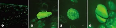
圖 1 正常軟骨Ⅵ型膠原IF染色及3D重建結果A: 正常全層軟骨; B ~ D: 正常軟骨細胞及Ⅵ型膠原的3D重建圖像 (B: 表面層; C: 移行層; D: 放射層)Fig. 1 IF staining and 3D images of type Ⅵ collagen in articular cartilage of healthy knees A: full thickness cartilage (40×); B-D: 3D images of chondrocyte and type Ⅵ collagen coat (B: surface layer; C: transition layer;D: radiation layer) (150-200 serial sections of 2 048×2 048 pixels at an interval of 0.2μm using DeltaVision with 60×objective lens. Chondrocyte was non-transparent while type Ⅵ collagen colored green and was transparent). Scale Bars:40μm, B:3μm, C:3μm, D:4μm

圖 2 正常軟骨和OA軟骨中軟骨細胞體積和Ⅵ型膠原體積測量結果 A: 正常軟骨(Vc、Vp); B: OA軟骨(Vc’、Vp’)Fig. 2 Volumn of chondrocyte (Vc, Vc’) and volumn of type Ⅵ collagen (Vp, Vp’) in each layer in healthy cartilage and OA cartilage A: healthy cartilage (aP<0.01,bP<0.01); B: OA cartilage (aP<0.01,bP<0.01)Note: Surf: surface layer; Trans: transition layer; Radi: radiation layer

圖 3 OA軟骨Ⅵ型膠原IF染色及3D重建結果A:全層OA軟骨;B ~ D:OA軟骨細胞及Ⅵ型膠原的3D重建圖像 (B:移行層單細胞; C:放射層成列細胞; D:放射層成團細胞)Fig. 3 IF staining and 3D images of type Ⅵ collagen in articular cartilage of OA knees A: full thickness cartilage (40×); B - D: 3D images of chondrocyte and type Ⅵ collagen coat; B: transition layer (left: fluorescent 3D images; right: surface reconstruction images corresponding to the left, chondrocyte colored yellow while type Ⅵ collagen colored green);C: 3 chondrocytes in row in radiation layer (left: fluorescent 3D image; right: surface reconstruction image, type Ⅵ collagen colored green); D: 5 chondrocytes in crowd in radiation layer (left: fluorescent 3D image; right: surface reconstruction image, type Ⅵ collagen colored green). Scale bars: A: 40μm; B: 2μm; C: 8μm; D: 7μm
骨關節炎是最常見的關節退行性疾病,關節軟骨表層的Ⅱ型膠原變性和蛋白多糖流失是OA的特征性病理改變[12-13],其形成的主要原因是軟骨細胞表型改變,即退變。Ⅵ型膠原與軟骨細胞關系密切,是OA軟骨細胞表型改變的可能原因。目前關于OA軟骨中Ⅵ型膠原的分布規律并未形成共識。Hambach等[14]報道了OA軟骨單位中Ⅵ型膠原含量較正常軟骨單位顯著增加;但Polur等[15]通過原位免疫標記觀察證實OA軟骨中Ⅵ型膠原在酶解作用下含量減少。這種分歧的產生可能是由于關節軟骨是非均一物質,從表面帶到深層的鈣化帶,無論細胞還是基質的形態和性質都有很大差異,基于酶解分離的軟骨細胞研究[14-16]或基于二維圖像的物質定量[15,17]都忽視了原位細胞和基質的空間結構,因此難以得到一致的結論。
Youn等[18]和Choi等[19]利用激光共聚焦顯微鏡斷層掃描圖像構建正常軟骨細胞及PCM的3D模型,實現了軟骨微觀結構的原位重現和定量分析。受之啟發,將軟骨組織在Delta-Vision Elite成像系統下進行3D觀察,并且利用其自帶的SoftWoRx軟件的還原型迭代去卷積模塊減少散射熒光的影響,使3D圖像分辨率和處理效果進一步提高。基于以上實驗方法的改進,本研究對正常軟骨和OA軟骨中Ⅵ型膠原進行了體積測量及空間分布的描述,結果顯示,OA軟骨中Ⅵ型膠原體積減小,形態不規則,PCM中的Ⅵ型膠原呈蜂窩狀,甚至出現大面積空洞,且空洞的位置無明顯規律。本實驗測得正常軟骨中,軟骨細胞體積與相關文獻相近,但各層細胞PCM中的Ⅵ型膠原厚度和體積均較小[18]。其主要原因是去卷積處理后,散射熒光被去除,熒光范圍減小;同時,Imaris軟件中進行表面重建時,設定的熒光閾值較小。以上兩種處理方法使3D圖像能更接近組織的真實情況,減小了Ⅵ型膠原厚度和體積測量的誤差。
此外,在放射層出現了無規則形狀的散在熒光,這些熒光距細胞甚遠,存在于PCM以外的基質中,可能是S?der等[4]報道的Ⅵ型膠原含量升高的主要原因。根據OA軟骨中軟骨細胞的表型改變和數量減少,可以推測其來源于軟骨細胞的異常分泌作用[16]或軟骨細胞凋亡[20]后殘留分散的Ⅵ型膠原。S?der等[4]曾描述過這種散在熒光,并且認為與正常軟骨中Ⅵ型與Ⅱ型膠原存在交聯不同,這些散在的Ⅵ型膠原與Ⅱ型膠原并無交際。因此,推測這些Ⅵ型膠原與正常軟骨細胞周基質中的Ⅵ型膠原有所不同,其理化性質有待進一步研究。
1 Zamli Z, Sharif M. Chondrocyte apoptosis: a cause or consequence of osteoarthritis?[J]. Int J Rheum Dis, 2011, 14(2):159-166.
2 Guilak F, Alexopoulos LG, Upton ML, et al. The pericellular matrix as a transducer of biomechanical and biochemical signals in articular cartilage[J]. Ann N Y Acad Sci, 2006, 1068:498-512.
3 Zhang ZJ, Jin W, Beckett J, et al. A proteomic approach for identification and localization of the pericellular components of chondrocytes[J]. Histochem Cell Biol, 2011, 136(2): 153-162.
4 S?der S, Hambach L, Lissner R, et al. Ultrastructural localization of type VI collagen in normal adult and osteoarthritic human articular cartilage[J]. Osteoarthritis Cartilage, 2002, 10(6):464-470.
5 Zelenski N, Leddy HA, Sanchez-Adamsj S, et al. CollagenVI: the Link between the extracellular matrix and chondrocyte mechanotranduction[J]. Trans Orthop Res Soc, 2014, 39: 192.
6 Alexopoulos LG, Youn I, Bonaldo P, et al. Developmental and osteoarthritic changes in Col6a1-knockout mice: biomechanics of type VI collagen in the cartilage pericellular matrix[J]. Arthritis Rheum, 2009, 60(3):771-779.
7 Pullig O, Weseloh G, Swoboda B. Expression of type VI collagen in normal and osteoarthritic human cartilage[J]. Osteoarthritis Cartilage, 1999, 7(2): 191-202.
8 Zamli Z, Adams MA, Tarlton JF. Increased chondrocyte apoptosis is associated with progression of osteoarthritis in spontaneous Guinea pig models of the disease[J]. Int J Mol Sci, 2013, 14(9): 17729-17743.
9 Muraoka T, Hagino H, Okano T, et al. Role of subchondral bone in osteoarthritis development: a comparative study of two strains of guinea pigs with and without spontaneously occurring osteoarthritis[J]. Arthritis Rheum, 2007, 56(10):3366-3374.
10 Ohlson MB, Huang ZW, Alto NM, et al. Structure and function of salmonella SifA indicate that its interactions with SKIP, SseJ, and RhoA family GTPases induce endosomal tubulation[J]. Cell Host Microbe, 2008, 4(5): 434-446.
11 Davis C-, Kim KY, Bushong EA, et al. Transcellular degradation of axonal mitochondria[J]. Proc Natl Acad Sci U S A, 2014, 111(26):9633-9638.
12 Loeser RF, Goldring SR, Scanzello CR, et al. Osteoarthritis: a disease of the joint as an organ[J]. Arthritis Rheum, 2012, 64(6):1697-1707.
13 Wilusz RE, Sanchez-Adams J, Guilak F. The structure and function of the pericellular matrix of articular cartilage[J]. Matrix Biol,2014, 39:25-32.
14 Hambach L, Neureiter D, Zeiler G, et al. Severe disturbance of the distribution and expression of type VI collagen chains in osteoarthritic articular cartilage[J]. Arthritis Rheum, 1998, 41(6):986-996.
15 Polur I, Lee PL, Servais JM, et al. Role of HTRA1, a serine protease, in the progression of articular cartilage degeneration[J]. Histol Histopathol, 2010, 25(5): 599-608.
16 Horikawa O, Nakajima H, Kikuchi T, et al. Distribution of type VI collagen in chondrocyte microenvironment: study of chondrons isolated from human normal and degenerative articular cartilage and cultured chondrocytes[J]. J Orthop Sci, 2004, 9(1): 29-36.
17 Quinn TM, Hunziker EB, Hauselmann HJ. Variation of cell and matrix morphologies in articular cartilage among locations in the adult human knee[J]. Osteoarthritis Cartilage, 2005, 13(8): 672-678.
18 Youn I, Choi JB, Cao L, et al. Zonal variations in the threedimensional morphology of the chondron measured in situ using confocal microscopy[J]. Osteoarthritis Cartilage, 2006, 14(9):889-897.
19 Choi JB, Youn I, Cao L, et al. Zonal changes in the threedimensional morphology of the chondron under compression:The relationship among cellular, pericellular, and extracellular deformation in articular cartilage[J]. J Biomech, 2007, 40(12):2596-2603.
20 Mobasheri A, Matta C, Zákány R, et al. Chondrosenescence:definition, hallmarks and potential role in the pathogenesis of osteoarthritis[J]. Maturitas, 2015, 80(3):237-244.
Changes of distribution of type Ⅵ collagen in osteoarthritis cartilage based on threedimensional images in situ
LI Zhao1,2, LI Peng1,2, WANG Hengsha3, CHANG Tongjie4, GAO Yingying4, CHEN Chao1, ZHANG Licheng1, ZHANG Lihai1,ZHANG Yi2, TANG Peifu1
1Chinese PLA General Hospital, Beijing 100853, China;2Department of Cell Biology, Institute of Basic Medical Sciences, Academy of Military Medical Sciences, Beijing 100850, China;3Soonbio Pathology Technology Laboratory, Beijing 100039, China;4Imaging Core Facility, Tsinghua University Branch of China National Center for Protein Sciences, Beijing 100084, China
Corresponding author: TANG Peifu. Email: pftang301@163.com; ZHANG Yi. Email: zhangyi612@hotmail.com
Objective To explore the changes of spatial distribution of typeⅥ collagen in osteoarthritis cartilage. Methods Frozen sections of full thick cartilage from osteoarthritis and healthy knee of guinea pig in mode group and control group were performed immunofluorescence staining, and then scanned by Delta-Vision Elite system. The scanning data were processed and analyzed by Imaris software, the volume of chondrocyte (Vc), thickness and volume of typeⅥ collagen (Vp) of the two groups were measured. Results In healthy cartilage, every chondrocyte wore a thin coat of type Ⅵ collagen, whose thickness had no significant difference(P>0.05), and the volume increased (P<0.01) with the increase of depth. In osteoarthritis cartilage, the volume of typeⅥcollagen coat decreased and distributed irregularly. It showed honeycomb distribution and scattered distribution in transition layer and radiation layer, respectively (P<0.01). Moreover, the scattered type Ⅵ collagen was found in extracellular matrix. Conclusion The irregular distribution of typeⅥ collagen in the osteoarthritis cartilage may be one of the factors which induces chondrocyte degeneration.
type Ⅵ collagen; chondrocyte; osteoarthritis; spontaneous guinea pig model of osteoarthritis
R 684.3
A
2095-5227(2015)07-0734-04
10.3969/j.issn.2095-5227.2015.07.025
2015-03-24
李曌,男,在讀碩士。研究方向:骨關節炎發病機制。Email: lizhao0215@163.com
唐佩福,男,主任醫師,博士生導師,骨科醫院院長。Email: pftang301@163.com;張毅,女,研究員,博士生導師,主任。Email: zhangyi612@hotmail.com
猜你喜歡
中老年保健(2021年5期)2021-12-02 15:48:21
中國民間療法(2021年18期)2021-11-02 08:20:18
中國民間療法(2021年14期)2021-08-30 08:24:36
中國民間療法(2021年12期)2021-07-28 07:31:16
中國民間療法(2021年5期)2021-06-09 09:21:18
基層中醫藥(2021年9期)2021-06-05 07:14:14
中國民間療法(2020年22期)2021-01-07 07:39:44
今日農業(2020年19期)2020-12-14 14:16:52
臨床醫藥文獻雜志(電子版)(2017年11期)2017-05-17 04:48:39
中國中醫藥現代遠程教育(2014年11期)2014-08-08 13:23:44