Calpain-1及Calpain-2在不同遷移能力肝癌細胞系中的表達*
2016-06-30 03:19:50陳騰祥劉振華王飛清
貴州醫科大學學報 2016年6期
關鍵詞:遷移能力
王 寧, 陳騰祥, 劉振華, 王飛清, 潘 婭
(1.貴州省骨科醫院 藥劑科, 貴州 貴陽 550002; 2.貴州醫科大學 生理教研室, 貴州 貴陽 500004; 3.貴州省人民醫院 肝膽外科, 貴州 貴陽 550001; 4.貴州省中醫院 檢驗科, 貴州 貴陽 550001)
Calpain-1及Calpain-2在不同遷移能力肝癌細胞系中的表達*
王寧1**, 陳騰祥2***, 劉振華3, 王飛清4, 潘婭2***
(1.貴州省骨科醫院 藥劑科, 貴州 貴陽550002; 2.貴州醫科大學 生理教研室, 貴州 貴陽500004; 3.貴州省人民醫院 肝膽外科, 貴州 貴陽550001; 4.貴州省中醫院 檢驗科, 貴州 貴陽550001)
[摘要]目的: 研究鈣激活中性蛋白酶蛋白-1(Calpain-1)及Calpain-2在不同遷移能力肝癌細胞系中的表達。方法: 常規細胞培養高遷移能力的MHCC97-H、低遷移能力的MHCC97-L、無遷移能力的HepG2的人肝癌細胞及正常人肝HL-7702細胞,采用western-bolt方法觀察Calpain-1及Calpain-2在3株人肝癌細胞及正常肝細胞中的表達。結果: Calpain-1在正常肝細胞HL-7702、高遷移能力的MHCC97-H、低遷移能力的MHCC97-L及無遷移能力的HepG2細胞中均有表達,表達量差異無統計學意義,P>0.05;與正常肝細胞HL-7702比較,高、低、遷移能力肝癌細胞系中Calpain-2不同程度表達,隨著細胞遷移能力增強而成遞增趨勢,P<0.01或P<0.05。結論: Calpain-2隨肝癌細胞遷移能力的增強表達增加,表明Calpain-2與肝癌細胞的侵襲轉移可能有密切關系。
[關鍵詞]癌,肝細胞; 鈣激活中性蛋白酶蛋白; 遷移能力
原發性肝癌(primary hepatocellular carcinoma,PHC)目前在我國的死亡率高居癌癥的第2位,據相關文獻調查其患病率約占全球癌癥患病率的55%[1-2]。研究發現,組織中的鈣激活中性蛋白酶(calcium activated neutral protease,Calpain)對某些腫瘤細胞的遷移能力有明顯影響[3],這在防治肝癌轉移靶點研究中報道甚少。本研究選擇不同轉移能力的肝癌細胞MHCC97-H、MHCC97-L、HepG2和正常肝細胞HL-7702作為研究對象,通過Western Blot研究Calpain家族中Calpain-1和Calpain-2蛋白在肝癌患者發生癌細胞轉移中的表達情況,為臨床治療肝癌發生癌細胞轉移提供新的治療靶點和基礎數據。
1材料與方法
1.1材料試劑與儀器
人肝癌細胞系MHCC97-H、MHCC97-L、HepG2和正常人肝細胞HL-7702均由中國科學院上海生命科學院細胞庫提供,DMEM培養基(Hyclone公司),胎牛血清(FBS),原裝引進USA Cell Signaling Technology(Calpain-1、2的一抗、羊抗兔二抗血清),原裝引進USA Merck Millipore PVDF轉化膜,Thermo Scientific CO2培養箱,實驗通用蛋白定量試劑盒(BCA)、碧云天生物技術公司產蛋白質上樣緩沖液,Beckman Coulter產實驗專用高速冷凍離心機, Syngene 產GBOX iChemiXR化學發光及凝膠成像儀,北京六一公司產垂直型蛋白電泳系統和電泳儀。
1.2方法
1.2.1細胞的培養與處理人肝癌細胞系MHCC97-H、MHCC97-L、HepG2和正常肝細胞HL-7702細胞為貼壁生長,用DMEM培養基+10% FBS(胎牛血清),放置在37 ℃,5% CO2孵箱環境中培養。當培養生長的癌細胞密度為80%左右時,對癌細胞進行計數,將癌細胞傳代與標記皿中繼續培養,在冰上用RIPA裂解液裂解培養已伸展完全的癌細胞,低溫高速離心細胞液取上清液制備蛋白樣品。
1.2.2免疫印記實驗 依據蛋白定量試劑盒實驗步驟,對樣品蛋白進行定量,實驗樣本化學發光顯色,用免疫印記實驗凝膠成像儀對已顯色的樣本成像顯示Calpain-1及Calpain-2 含量和磷酸化情況分析。
2結果
2.1Calpain-1表達
Western blot檢測發現,不同轉移能力的人肝癌細胞(MHCC97-H、MHCC97-L、HepG2)中Calpain-1蛋白表達,與正常人肝細胞HL-7702相比,Calpain-1的表達變化不明顯,P>0.05。見圖1。
2.2Calpian-2表達
如圖2所示,Calpain-2在3種肝癌細胞中的表達量明顯高于正常肝細胞HL-7702 。與正常肝細胞HL-7702比較,高轉移肝癌細胞MHCC97-H中 Calpain-2的表達增高明顯,P<0.01,其低分子量(low molecular weight,LMW)剪切片段形式顯著增多,可見Calpain-2被剪切有至少6種LMW片段;在低轉移肝癌細胞MHCC97-L中,Calpain-2的表達異常,P<0.01,Calpain-2也可被剪切2種LWM片段;不轉移的肝癌細胞(HepG2)Calpain-2的表達比HL-7702高,P<0.05。
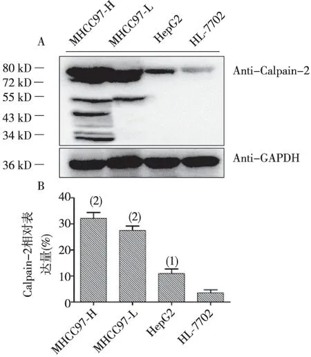
注:A為Western blot結果,B為A的統計結果(1)P<0.05,(2)P<0.01 vs Control圖2 MHCC97-H、MHCC97-L、HepG2和HL-7702中Calpain-2的表達Fig.2 Expressions of Calpain-1 in MHCC97-H,MHCC97-L, HepG2 and, HL-7702
3討論
腫瘤發生發展是多因素、多基因、多階段才最終形成的十分復雜的生物學現象,具有細胞黏附、基質降解、細胞運動3個最基本步驟。Calpain是鈣蛋白Cal(Calmoclulin)和木瓜蛋白酶pain(Papain)兩者以非共價鍵結合形成。Calpain有多種同工酶,按照其分布特點可將其分為兩大類,一類具有組織特異性,另一類非組織特異性蛋白酶[4]。近年來,非組織特異性蛋白酶的研究比較深入,其已知的2種亞型是Calpain-1(也稱μ-calpain)和Calpain-2(也稱m-calpain),它們的激活需要不同濃度的鈣離子水平,在不同的生理或病理狀態下,Calpain-1或Calpain-2的生物學作用也不同[5],在很多應激的情況下,Calpain家族蛋白的表達和活性都會發生相應的變化,而這些活性的變化與細胞周期、細胞信號轉導及細胞增殖、分化及細胞凋亡[6]、細胞骨架重塑與遷移有著密切的關系,在癌細胞中,這種明顯異常的變化,提示Calapin家族蛋白參與了癌細胞的惡性演變,研究認為Calpain與細胞的遷移浸潤有關[7-8]。已有研究發現乳腺癌或前列腺癌細胞中,Calpain-2可以直接使細胞遷移方向的后方細胞皮質的絲狀肌(filamentousactin,F-actin)解體[9]。相關文獻前期研究報道,癌細胞胞內鈣離子濃度在惡性腫瘤細胞中的含量表現明顯的升高,其細胞的離子內環境平衡狀態發生紊亂,實驗研究提示肝癌細胞中的Calpain 發揮功能機制作用可能是在高濃度Ca2+的環境下[10]。腫瘤的侵襲和轉移是導致腫瘤治療效果不佳和患者最終出現死亡的重要原因之一,也是極為復雜、多分子水平改變、涉及多基因的過程,大約有細胞異常的增殖、腫瘤細胞從原發灶上脫落、侵犯組織血管或淋巴管、遷移,黏附著于遠處管腔床、侵入靶器官和遠處部位形成繼發腫瘤等表現[11]。而在以人纖維肉瘤細胞、膠質母細胞瘤細胞、乳腺癌細胞、前列腺癌細胞等癌細胞為基礎的實驗研究報道中,認為Calpain-2可以作為抑制腫瘤侵襲轉移的一個潛在靶點[12-16]。
本研究發現在肝癌細胞MHCC97-H、MHCC97-L、HepG2中Calpain的家族成員Calpain-2的表達顯著增多,尤其是在高轉移能力的肝癌細胞MHCC97-H中,calpain-2的蛋白表達最高,且隨著肝癌細胞遷移能力的增強,calipain-2的蛋白表達也呈現遞增現象,這一實驗結果提示Calpain-2在肝癌細胞的侵襲和遷移能力中發揮作用。
綜上所述,Calpain-2在3種肝癌細胞(無、低、高遷移能力)中的表達隨遷移能力的增強而增加,表明Calpain-2可能與肝癌的轉移、復發密切相關,其可能成為評價肝癌生物學行為的重要指標之一。Calpain-2有望成為肝細胞癌臨床靶向治療的新靶點,但其具體的作用機制還有待進一步研究。
4參考文獻
[1] Jemal A,Bray F,center MM,et al.Global cancer statistics [J].CA:a Cancer Journal for Clinicians, 2011(2):69-90.
[2] Center MM,Jenmal A.International trends in liver cancer incidence rates [J].Cancer Epidemiology Biomarkers & Prevention, 2011(11):2362-2368.
[3] Storr SJ,Carragher NO,Frame MC,et al. The calpain system and cancer[J].Nat Rev Cancer, 2011(5):364-374.
[4] Schád E,Farkas A,Jékely G,et al.A novel human small subunit of calpains. Biochem J, 2002(362):383-388.
[5] Goll DE,Thompson VF,Li H,et al.The calpain system[J].Physiol Rev, 2003 (3):731-801.
[6]Goll DE.The calpain system[J].Physiol Rev, 2003(3):731-801.
[7] Jang HS,Lal S,Greenwood JA.Calpain 2 is required forglioblastoma cell invasion: regulation of matrix metalloproteinase 2[J].Neurochem Res, 2010 (11):1796-1804.
[8] Wu M,Yu Z,Fan J,et al.Functional dissection of human protease mu- calpain in cell migration using RNAi[J].FEBS Lett, 2006(13):3246-3256.
[9] Storr SJ,Lee KW,Woolston CM,et al.Calpain system protein expression in basal-like and triple-negative invasive breast cancer[J].Ann Oncol, 2012(9):2289-2296.
[10]Tang J, Wu YM, Zhao P, et al. Overexpression of HAb18G/CD147 promotes invasion and metastasis via alpha3beta1 integrinmediated FAK-paxillin and FAK-PI3K-Ca2+pathways [J]. Cell Mol Life Sci, 2008(18):2933-2942.
[11]Nakayama H,Yasui W, Yokozaki H,et al.Reduced expression of nm23 is associated with metastasis of human gastric carcinomas[J].Jpn J Cancer Res, 1993(2):184-190.
[12]Jang HS,Lal S,Greenwood JA.Calpain 2 is required for glioblastoma cell invasion: regulation of matrix metallopro-teinase 2[J].Neurochem Res, 2010 (11):1796-1804
[13]Carragher NO,Walker SM,Scott Carragher LA,et al.Calpain 2 and Src dependence distinguishes mesenchymal and amoeboid modes of tumour cell invasion: a link to integrin function[J].Oncogene, 2006(42):5726-5740.
[14]Mamoune A,Luo JH,Lauffenburger DA,et al.Calpain-2 as a target for limiting prostate cancer invasion[J].Cancer Res, 2003(15):4632-4640.
[15]Noma H,Kato T,Fujita H,et al.Calpain inhibition induces activation of the distinct signalling pathways and cell migration in human monocytes[J].Immunology, 2009 (1):e487-e496.
[16]Storr SJ,Carragher NO,Frame MC,et al.The calpain system and cancer[J].Nat Rev Cancer, 2011(5):364-374.
(2016-03-12收稿,2016-05-28修回)
中文編輯: 潘婭; 英文編輯: 劉華
Expression of Calpain 1 and 2 in Liver Cancer Cells with Different Migration Abilities
WANG Ning1, CHEN Tengxiang2, LIU Zhenhua3, WANG Feiqing4, PAN Ya2
(1.DepartmentofPharmaceuticalPreparation,GuizhouProvinceOstelolgicalHospital,Guiyang550002,Guizhou,China;2.DepartmentofPhysiology,GuizhouMedicalUniversity,Guiyang550004,Guizhou,China; 3.DepatmentofLiverandGallSurgery,GuizhouProvincialPeople'sHospital,Guiyang550002,Guizhou,China; 4.ClinicalLaboratory,GuizhouProvincialHospitalofTraditionalChineseMedicine,Guiyang550001,Guizhou,China)
[Abstract]Objective: To investigate protein expression of Calpain-1 and Calpain-2 in liver cancer cell line with different migration ability. Methods: MHCC97-H (highly migratory liver cancer cells), MHCC97-L (lowly migratory liver cancer cells), HepG2 (liver cancer cells with no migration ability) and HL-7702 (normal liver cells) were routinely cultured. Western-blot method was used to observe protein expression of Calpain-1 and Calpain-2 in the three human liver cancer cell lines and the normal liver cell. Results: There were protein expressions of Calpain-1 in HL-7702, MHCC97-H, MHCC97-L and HepG2, and the difference was not statistically significant (P<0.05). Compared with normal liver cells HL-7702, Calpain-2 showed the different degrees of protein expression in high, low, migration ability of liver cancer cell line , and the protein expression of Calpain-2 significantly enhanced with increasing cell migration ability (P<0.01 or P<0.05). Conclusions: Calpain-2 shows an increasing protein expression with the enhancement of the migration ability of liver cancer cells, which indicates that calpain-2 may be closely related to the invasion and metastasis of liver cancer cells.
[Key words]carcinoma,hepatocellular; Calpain; migration ability
*[基金項目]貴州省科技廳科研基金[黔科合LG字(2011)008]; 貴州省教育廳自然科學研究項目[黔教科(2008)022]
[中圖分類號]R735.7
[文獻標識碼]A
[文章編號]1000-2707(2016)06-0649-04
DOI:10.19367/j.cnki.1000-2707.2016.06.007
**貴州醫科大學2014屆碩士研究生
***通信作者 E-mail:710232517@qq.com;371251826@qq.com
網絡出版時間:2016-06-16網絡出版地址:http://www.cnki.net/kcms/detail/52.5012.R.20160616.1742.058.html
猜你喜歡
中國科技博覽(2017年2期)2017-03-30 19:23:34
新課程·中旬(2017年1期)2017-03-27 21:13:22
數學學習與研究(2017年4期)2017-03-20 11:45:08
新課程·上旬(2016年10期)2017-03-20 07:04:22
讀與寫·教育教學版(2017年3期)2017-03-20 02:46:02
知識窗·教師版(2017年1期)2017-03-15 08:49:12
成長·讀寫月刊(2016年11期)2016-12-14 22:42:57
求知導刊(2016年18期)2016-08-10 18:14:19
人民論壇(2016年5期)2016-03-24 22:13:11
中學課程輔導·教師通訊(2015年18期)2015-11-09 18:36:21
