Prevalence of focal lamina cribrosa defects in eyes with pachychoroid disease spectrum
2022-01-20 07:03:46HaeMinKangNaEunLeeJeongHoonChoiHyoungJunKohSungChulLee
關鍵詞:挑戰
INTRODUCTION
Pachychoroid disease spectrum (PDS) is a recently defined clinical entities characterized by retinal pigment epithelium (RPE) abnormalities overlying areas of choroidal thickening. PDS occurs due to pachychoroid-driven processes involving choroidal congestion and choroidal hyperpermeability, manifested in choroidal thickening and dilated choroidal vessels. The current definition of PDS includes central serous chorioretinopathy (CSC), pachychoroid pigment epitheliopathy (PPE), pachychoroid neovasculopathy,and polypoidal choroidal vasculopathy (PCV).
A number of studies have shown disease characteristics,suitable treatment strategies, and possible pathogenesis of PDS, however, still various aspects of PDS such as optic nerve head (ONH) structures remain to be uncovered. A few studies have presented ONH structures in the PDS. One study demonstrated that lamina cribrosa (LC) disinsertion or LC defect is associated with peripapillary retinoschisis in PDS as well as with glaucoma. Increased leakage from the hyperpermeable choroidal vessels may lead to greater inflow of fluid to the ONH from the subarachnoid space through peripheral LC disinsertion or from the vitreous region through central LC defects in eyes with PDS; however, the study only included cases with PDS having peripapillary retinoschisis. Recently, one study defined peripapillary pachychoroid syndrome (PPS)as a distinct variant of PDS,in which peripapillary choroidal thickening is associated with intraretinal and/or subretinal fluid in the nasal macula and occasional ONH edema. In that study, all eyes showed intraretinal fluid and cysts extending from the temporal disc margin, with associated atrophy of the RPE, photoreceptor ellipsoid zone, and external limiting membrane. However,they suggested that peripapillary intraretinal fluid resembling peripapillary retinoschisis can occur in the eyes with PDS that do not show LC disinsertion.
許多年了,這一次(也是唯一一次)榮耀地接受采訪,我清晰地記住了那個女記者。那天女記者穿著一件淡紅的短袖上衣,兩個乳房驕傲又堅挺,白色的短擺裙子下露出了白如蘿卜的細腿,還有一頭烏黑發亮的頭發,清風吹起幾縷覆了她的眼、眉和臉頰兩側的紅潤后,隨即她就用白玉似的手指掠到了耳后。她的表情滿是矜持與憐憫,眼神直直地盯著我們。他媽的,這個女記者真是美,實在太漂亮太迷人了。事后,我每次撒尿的時候心里就會想起她:如果我娶媳婦能娶上這個女記者樣的女人,那該有多好啊。
Despite several studies have suggested the possible correlation of LC disinsertion/defect in the patients with PDS, there has been no study investigating the prevalence of focal LC defect among the patients with PDS. Based on these studies,we intended to examine the prevalence of focal LC defect among the patients with pachychoroid diseases, but without peripapillary retinoschisis. We also compared the prevalence of focal LC defects between the eyes with PDS in the absence of peripapillary retinoschisis and that of the normal control group.
SUBJECTS AND METHODS
This study was approved by the Institutional Review Board of International St. Mary’s Hospital and adhered to the tenets of the Declaration of Helsinki. Due to the retrospective nature of the study, the requirement for informed consent was waived.
仿制藥企業所提的專利挑戰中,大多數針對的是非活性成分專利。極高的專利挑戰成功率激勵更多的仿制藥企業對非活性成分專利提出專利挑戰。而從原研藥企業角度而言,非活性成分專利極高的挑戰成功率又“迫使”其在桔皮書登記更多的專利,從而通過數量的優勢來彌補非活性成分專利在保護效果上的不足。桔皮書中更多的非活性成分專利又必然會導致更多的針對非活性成分專利的挑戰。這樣就形成了一個“怪圈”,使得雙方都需要付出更多的精力和資源來維護或挑戰改進型的非活性成分專利。
Three-dimensional computed tomography reconstruction indicating a rich collateral renal blood supply and confirming the origins and extent of the renal artery stenosis.
However, based on our findings, we discarded the primary hypothesis that increased hydrostatic pressure of PDS may be associated with development of LC disinsertion/defect. At least the eyes with PDS but without peripapillary retinoschisis,it seems that increased hydrostatic pressure does not have significant impact on the development of LC alteration.Simply, the presence of focal LC defect among the patients with PDS may be just accidental findings of normal ONH variants in this study.Previously, our study group investigated the prevalence of focal LC defects in the patients with unilateral branch retinal vein occlusion (BRVO), those with normal tension glaucoma(NTG), and the normal control group. In that study,prevalence of focal LC defects were significantly higher in the eyes with BRVO and those with NTG than that of the normal control group: the prevalence of focal LC defects was 38.9%in the BRVO group, 41.7% in the NTG group, and none in the control group. In addition, the mean peripapillary choroidal thickness was significantly thinner in the eyes with focal LC defects than those without in both the BRVO group and the NTG group. Based on these results, our study group assumes the possible pathophysiologic correlation between BRVO and NTG. Although this study lacks of the NTG group, we could infer that focal LC defects in the patients with PDS may not be significantly correlated with pathologic changes of ONH,because there was no significant difference in the prevalence of focal LC defects between the PDS group and the normal control group.
Our study had several limitations, including a relatively small study population. Moreover, due to the retrospective nature of the study, the control group was relatively heterogeneous.Further investigations with larger populations, a longitudinal design should enhance our understanding of LC disinsertion in patients with PDS.
Ophthalmologic examination was performed for each patient according to our clinic’s standard retinochoroidal evaluation procedure, as described previously. Routine ophthalmologic evaluation included slit lamp examination, intraocular pressure measurement using a non-contact tonometer, and fundus examination after dilation. The refractive error was measured for each eye using an autorefractor followed by conversion to the spherical equivalent [diopters (D)]. For the diagnosis of CSC, PCV, and pachychoroidal neovasculopathy, fluorescein angiography and indocyanine green angiography were conducted with a Heidelberg Retina Angiograph System (HRA-2; Heidelberg Engineering, Heidelberg, Germany) with a confocal scanning laser ophthalmoscope.
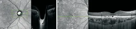
Spectral-domain optical coherence tomography (SD-OCT; Spectralis/Heidelberg Engineering)with EDI modality was used to examine the ONH and macula. For LC analysis, serial horizontal cross-sectional scans, approximately 30 μm apart and coering the ONH, were obtained (Figure 1A). EDI-OCT images of the ONH were reviewed by experienced glaucoma specialist (Lee NE), who was blinded to the retinal status; the presence of alterations in the LC was evaluated. Focal LC defect was defined as an anterior laminar surface irregularity violating the normal smooth curvilinear U- or W-shaped contour. To avoid falsepositives, defects needed to be >100 μm in diameter, >30 μm in depth, and present in at least one additional raster scan, as suggested by a previous study.
The mean subfoveal choroidal thickness (SFCT) was also measured using the Spectralis SD-OCT system with EDI.Choroidal thickness was defined as the perpendicular distance between the outer border of the hyperreflective line corresponding to the RPE, and the chorio-scleral interface.For macular evaluation, serial cross-sectional horizontal scans were obtained, approximately 121 μm apart in a 30°×15°macular area. Single horizontal and vertical scans across the fovea were acquired separately. To measure SFCT, at least two horizontal and vertical high-quality scans throughout the fovea were obtained from each eye. Utilizing the digital calipers in the Heidelberg Spectralis OCT software, the choroidal thickness was measured horizontally and vertically at the subfoveal region in each trans-sectional image, and the average measurement was calculated (Figure 1B). Three CharacteristicsPatients with PDS (=180)Patients without PDS (=236)independent observers (Kang HM, Lee NE, and Choi JH)blinded to the clinical data (including the ONH status of each patient) measured the choroidal thicknesses.For statistical analysis, eyes with PDS, including CSC, PCV,and pachychoroid neovasculopathy, were included in the study.In cases with bilateral PDS eyes, the right eye was analyzed.
分析工具 本研究采用CiteSpace 5.0對數據進行分析:從CNKI下載檢索數據并在CiteSpace中進行數據轉換,導入數據并進行參數設置,運行軟件進行圖譜繪制。
RESULTS
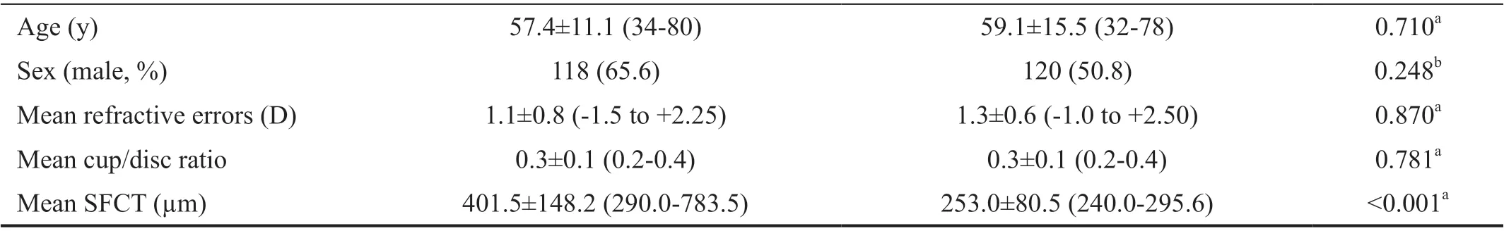
We retrospectively reviewed 180 consecutive patients with PDS and 236 patients without PDS, who met the study criteria.Among the patients with PDS, 118 (65.6%) were male and the mean age at the time of examination was 57.4±11.1y (range:34-80y). There were no significant differences in clinical characteristics between the patients with PDS and the control group, including age (=0.710) and sex (=0.248). The mean SFCT was significantly higher in the PDS group than in the control group (<0.001). A detailed comparison of the clinical characteristics between the two groups is shown in Table 1.Among the patients with PDS, the primary diagnosis was CSC in 73 (40.6%), PCV in 75 (41.7%), and pachychoroidal neovasculopathy in 32 (17.8%); all patients underwent intravitreal anti-vascular endothelial growth factor injections.
Among 180 patients with PDS, 6 (3.3%) showed focal LC defects in the absence of peripapillary retinoschisis. No patients in the control group showed focal LC defect. Diabetic retinopathy(DR) was not observed in any patient. The prevalence of focal LC defect was not significantly different between patients with and without PDS (=0.318). Among the six patients with PDS and focal LC defects, four eyes had PCV, one eye was the fellow eye of the PCV eye, and one eye showed pachychoroidal neovasculopathy. The detailed characteristics of the six patients with focal LC defects are listed in Table 2,and representative images are shown in Figures 2-6.
目前,由于一維混沌映射其產生方式簡單,生成序列眾多而被廣泛應用于擴頻通信中,現在比較常見的、統計性能較好的一維混沌映射主要有以下3種,其表達式以及初值和系統參數的取值范圍如下。
登錄APP查看全文
猜你喜歡
小哥白尼(趣味科學)(2021年7期)2021-11-05 07:25:38
小哥白尼(野生動物)(2021年2期)2021-07-16 08:35:28
童話世界(2020年32期)2020-12-25 02:59:22
童話世界(2020年26期)2020-10-27 02:23:44
中國外匯(2019年15期)2019-10-14 01:00:36
動漫星空(興趣英語)(2019年3期)2019-03-06 01:55:00
專用汽車(2016年8期)2016-03-01 04:16:05
就業與保障(2015年9期)2015-04-17 03:41:47
 International Journal of Ophthalmology
2022年1期
International Journal of Ophthalmology
2022年1期
- International Journal of Ophthalmology的其它文章
- lnstructions for Authors
- Comment on: Trends in research related to high myopia from 2010 to 2019: a bibliometric and knowledge mapping analysis
- Progress of clinical therapies for dry age-related macular degeneration
- Observation seasonal variation of intraocular pressure in young healthy volunteers
- Effectiveness of oral probiotics supplementation in the treatment of adult small chalazion
- Characterization and validation of a chronic retinal neovascularization rabbit model by evaluating the efficacy of anti-angiogenic and anti-inflammatory drugs