Heparanase-1 is downregulated in chemoradiotherapy orbital rhabdomyosarcoma and relates with tumor growth as well as angiogenesis
2022-01-20 07:00:34WeiQiangTangYanHeiJingLin
INTRODUCTION
R habdomyosarcoma (RMS) is a rare type of soft tissue sarcoma with a high degree of malignancy and mortality.Head and neck RMS occur frequently, and approximately 25%-30% of them appear in the orbit. It is well known as a pediatric disease. In adults, its incidence is pretty low,and prognosis is significantly worse, compared with those in children. RMS are subdivided into 4 types in the 2013 WHO classification, including embryonal, alveolar,spindle cell/sclerosing, and pleomorphic RMS. Embryonal rhabdomyosarcoma (ERMS) and alveolar rhabdomyosarcoma(ARMS) were the primary histologies. In addition to differences in clinical presentations and outcomes, a number of genetic features separate ARMS from ERMS. The alveolar type is less frequent with a worse prognosis. Therefore,histology was considered to be a major treatment determinant and the most important prognostic factor. The commonest embryonal type is significantly correlated with orbital involvement. Excellent prognosis of orbital RMS can be achieved frequently by treatment based on combination chemotherapy together with local surgery therapy and/or radiation therapy. However, local complications are common, including cataract, keratopathy, orbital hypoplasia or fat atrophy, eyelid malposition and lacrimal duct stenosis.To minimize ophthalmic adverse effects, novel therapeutic strategies are urgently needed for this malignancy. There are several attractive therapeutic strategies that have been investigated in RMS to date, including those directed against receptor tyrosine kinases and associated downstream signaling pathways, the Hedgehog signaling pathway, apoptosis pathway,DNA damage response, cell-cycle regulators, oncogenic fusion proteins, and epigenetic modifiers. Our previous study showed that there is a parallel between over-expression of heparanase-1 (HPSE-1) and key molecules of the Hedgehog signaling pathway in untreated human alveolar orbital RMS.HPSE‐1 is the only known mammalian endo‐β‐glucuronidase that can decompose heparan sulfate proteoglycans (HPSG)within the extracellular matrix (ECM), basement membrane(BM) or on the cellular surface. Its activity has been strongly implicated in cell invasion and migration—a consequence of the structural modification that loosens the ECM barrier, thereby it is involved in the regulation of multiple cellular processes and biological activities. Unlike matrix metalloproteinases (MMPs) enzyme families, HPSE-1 and its only close homolog HPSE-2 exert opposing biological properties. Previous studies have confirmed that elevated expression of HPSE-1 dramatically enhances tumor growth,angiogenesis, and metastasis, and it can be served as an independent prognostic factor for poor outcome of some cancer patients. The heparan sulfate mimetic PI-88 is a complex mixture of sulfated oligosaccharides that was identified as a potent inhibitor of HPSE-1 and subsequently entered clinical trials for cancer. It progressed to Phase III trials but ultimately was not approved for use because of the antibodyinduced thrombocytopenia in some patients. Advances in the chemistry of the heparan sulfate mimetics PG500 series provide numerous advantages over PI-88. PG545 has been selected as the lead clinical candidate for oncology and is currently undergoing formal preclinical development as a novel treatment for advanced cancer.
Data were collected and expressed as the mean±standard error (SD) and were analysed using SPSS statistical software (Ver 11.0, Chicago, IL, USA) with differences between groups assessed by Student’stest or one‐way analysis of variance (ANOVA).<0.05 was considered significant.
MATERIALS AND METHODS
The use of specimens from human subjects was approved by the Institutional Review Board of Chinese PLA General Hospital, and a signed consent form was obtained from each patient.
Formalin-fixed paraffin-embedded RMS specimens were obtained from 51 patients with orbital RMS,who were aged between 1 and 41y, and surgically treated in the Department of Ophthalmology, the Third Medical Centre,Chinese PLA General Hospital. Among these 51 patients, there were 28 ERMS and 23 ARMS; 27 cases (14 of ERMS and 13 of ARMS) received preoperative chemoradiotherapy followed by surgery (chemotherapy drugs include cyclophosphamide 100-400 mg, vincristine 1-1.5 mg and adriamycin 10-30 mg;The radiation dose is 60 Gy), while the other 24 patients received no such treatment before surgery.
The presence of HPSE-1 protein expression in 28 ERMS and 23 ARMS samples was confirmed by immunohistochemistry. The paraffin embedded tissues were sectioned with 3 μm thick and collected on glass slides, deparaffinized in xylene, and were then rehydrated in a graded ethanol series and washed in phosphate-buffered saline (PBS). The sections were immersed in citrate buffer(pH 6.0), and samples were heated in a pressure cooker for 10min to achieve antigen retrieval of HPSE-1. The rabbit anti-HPSE-1 (1:100; Abcam Ltd., Hong Kong, China.)was used as primary antibody. Sections were reacted with primary antibodies overnight at 4°C and washed three times in PBS, then followed by incubation with secondary antirabbit IgG polyclonal antibodies (1:250, Abcam) conjugated to horseradish peroxidase (HRP) for 30min at room temperature. A diaminobenzine (DAB) solution was added at room temperature for 3-5min to illuminate the positive staining signals, and the sections were counterstained with haematoxylin for 20s. Immunostaining results were assessed for intensity and extent by three experienced pathologists.Positive expression was defined as >10% of the cells showing moderate or intense positive cell staining by the antibody in 10 randomly selected fields on every section or >50% of the cells showing weak staining.
RD cells (a human RMS cell line), 293T cells and HUVECs were obtained from the cell bank of the Chinese Academy of Science (Shanghai, China) and incubated at 37°C in a humidified atmosphere which was maintained at 5% CO.
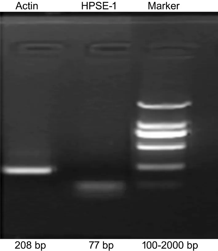
Four coding regions in the sequence of human HPSE mRNA (NM_006665.5) were designed as the target sequences. Four pairs of complementary DNA oligos were synthesized and annealed to generate a double-stranded oligo, followed by ligating into the linearized pcDNA6.2-GW/EmGFP-miR vector (Invitrogen, Carlsbad, CA, USA) using T4 DNA ligase at 22°C for 2h. 293T cells with high transfection efficiency of pcDNA6.2-EmGFP negative control plasmid were selected for plasmid transfection. For the relatively low abundance of HPSE-1 gene expression in 293T cells detected by reverse transcription polymerase chain reaction (RT-PCR; Figure 1), we cloned HPSE-1 cDNA into a pEGFP-N1 eukaryotic expression vector (Invitrogen)to construct a HPSE-1 eukaryotic plasmid. Using POLO deliverer? 3000 (Shanghai R&S Biotechnology Co., Ltd.,Shanghai, China) according to the manufacturer’s instruction,the HPSE-1 eukaryotic plasmid was then co-transfected with the recombinant vectors with HPSE-1 shRNA1, HPSE-1 shRNA2,HPSE-1 shRNA3, HPSE-1 shRNA4 (targeting different sequences of HPSE-1) or the Neg-shRNA into 293T cells,respectively. HPSE-1 mRNA and protein expressions were detected by RT‐PCR and Western blotting to filter out the most effective vector with HPSE‐1 shRNA.
RD cells and 293T cells were cultured in a mixture containing Dulbecco’s modified Eagle’s medium (DMEM; Invitrogen,Gaithersburg, MD) supplemented with 100 IU/mL penicillin and 100 μg/mL streptomycin, 1% GlutaMAX and 10% fetal bovine serum (FBS). HUVECs were cultured on ScienCellECM supplemented with 5% FBS, 100 U/mL penicillin,100 μg/mL streptomycin, 100 μg/mL endothelial cell growth factor.
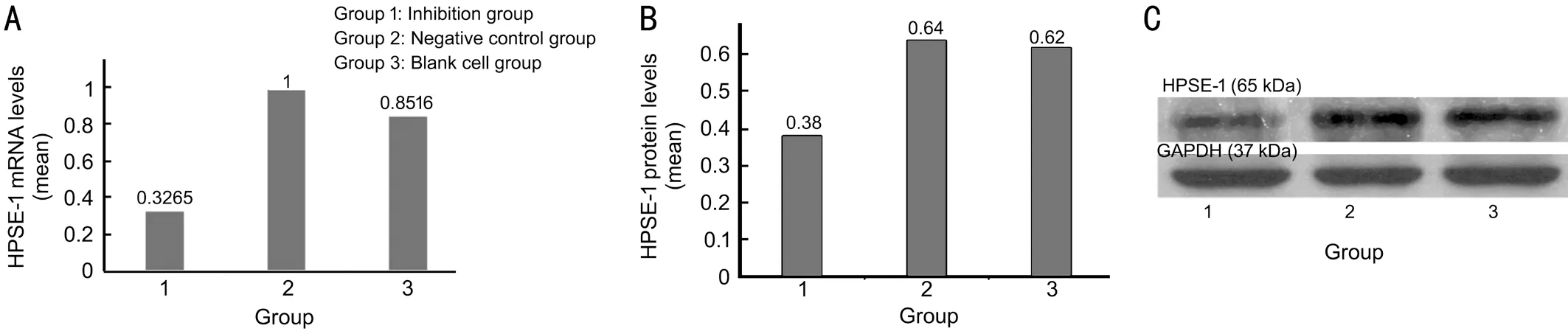
The HPSE-1 shRNA1 was used as the foundation for construction of the pLenti6.3-HPSE-1 RNAi lentivirus vector. Lentiviral vectors was packaged in 293T cells and the GFP expression was observed 72h post-package under the fluorescence microscope, the viral titer was calculated to be 2.75×10TU/mL. Recombinant lentiviruses with HPSE-1 shRNA were transduced into RD cells and the levels of HPSE-1 mRNA and protein were tested by real-time RT-qPCR and Western blotting in three groups of RD cells (inhibition group,negative control group, and blank cell group). As shown in Figure 4, HPSE-1 mRNA and protein expressions in the inhibition group were decreased by 67% and 41% respectively,when compared with the negative control group (<0.05). The lentivirus-mediated HPSE-1 RNAi notably down-regulated HPSE-1 expression levels of both mRNA and protein in RD cells.
Sequences of the most effective HPSE‐1 shRNA were inserted into the pLenti6.3/V5 DESTlentivirus RNAi expression system (Invitrogen, Carlsbad, CA, USA) to construct the HPSE-1 shRNA lentivirus vector. The insert was cloned into the pLenti6.3/V5 DEST plasmid by double digesting with AscI/PmeI enzymes and ligating with T4 DNA ligase. The resulting pLenti6.3-HPSE-1 shRNA was then sequenced and co-transfected with the ViraPower? Lentiviral Packaging Mix(Invitrogen) using POLO deliverer? 3000 (Shanghai R&S Biotechnology Co., Ltd.) into 293T cells. Viral supernatant was harvested 72h post-transfection and titers were determined by the green fluorescent protein (GFP) assay of the 293T cells infected with serial dilutions of concentrated lentivirus.
控制廢酸原液與水流量流比1∶1,考查廢酸酸度對酸回收效果的影響,試驗結果見表5所示。由表5可知,酸度提高對硫酸回收率及銅截留率影響不大。廢酸含酸在100~180g/L范圍,酸回收率均可保持在90%以上,銅截留率在85%以上。
RMS cells were randomly divided into 3 different groups,which were transfected with HPSE-1 shRNA (Inhibition group), Neg-shRNA (Negative control group), or untreated(blank cell group). The recombinant lentiviruses with HPSE-1 shRNA were transduced into RD cells with an optimized multiplicity of infection (MOI=10). Appropriate amount of HPSE-1 shRNA lentiviruses (2.745×10TU/mL)and negative control lentiviruses (2.0×10TU/mL) were added into the cells, 4‐8 μg/mL polybrene was also added to enhance the infection. The expression of lentivirus GFP was observed under fluorescence microscope 72h after infection and the percentage of fluorescent cells was more than 85%, which can be used for subsequent experiments.
我曾寫過一篇《鼓搗雜文之“三樂”》的小文,刊登在一家雜志上。說的是,寫作雜文有三樂:稿子出手,吐出胸中塊壘,一樂也;發表了,被社會認可,二樂也;稿費寄來,多少還有點勞動所得,三樂也。其實,寫雜文離不開讀雜文,而讀雜文,與寫雜文一樣,也有“三樂”。
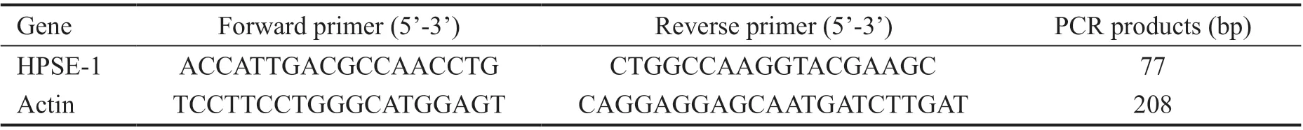
Total RNA was extracted from RD cells using Trizol (Invitrogen). The concentration and purity of RNA was subjected for a spectrophotometer analysis(Fullertone, CA, USA). RNA was reversely transcribed using M-MLV reverse transcriptase (Fermentas Inc., Burlington,Canada) at a total volume of 20 μL according to the manufacturer’s instructions. Subsequently, the qPCR assay was carried out using 2×SYBR Green qPCR Mix (Invitrogen)at the real‐time PCR amplification equipment of mastercycler EP realplex (Eppendorf AG, Hamburg, Germany). The PCR primers used to detect HPSE‐1 and β‐actin mRNA expression were seen in Table 1. PCR conditions included an initial step of 95℃ for 2min, followed by 40 cycles of 95℃ for 20s and then annealed at 60℃ for 15s, and 72℃ for 20s. Products were identified by melting curve analysis after 39 cycles.The comparative Ct (Ct) method was used to calculate the relative quantitation of mRNA as previously described. The expression of HPSE-1 was determined by normalization of the threshold cycle (Ct) of HPSE‐1 gene to that of β‐actin. The ΔCt and ΔΔCt were determined using the following equation:ΔCt = (Ct of HPSE‐1) ‐ (Ct of β‐actin), ΔΔCt = (mean ΔCt of HPSE‐1 genes in RNAi groups) ‐ (ΔCt of HPSE‐1 genes in control group). The 2implicates the relative expression vaule of the HPSE-1 gene.
反映其成礦是在基性火山熔巖噴發間歇期沉積形成,其圍巖為基性火山巖(斜長角閃巖)。說明其成礦物質來源除部分來自陸源外,大部分與其中火山沉積建造有關。
The proliferation of RD cell lines was evaluated by cell counting kit-8 (CCK-8) assay. RD Cells were seeded in 96-well culture plates at a density of 4000 cells per well. After the cells were transfected with the recombinant lentivirus at an MOI of 10 for 12h, the medium was replaced with a fresh complete medium.Blank cells and silenced cells were tested as well as the relative negative controls. After 24h incubation, 10 μL of CCK‐8 solution was added to each well for an additional 3h at 37℃.The number of proliferating cells was evaluated by measuring the absorbance at a 450 nm wavelength on a microtiter plate reader (Thermo Fisher Scientific, MA, USA) at the same time of 6 continuous days, and cellular viability was evaluated by the Avalue.
After washed two times with PBS at room temperature, the protein was extracted from RD cells using cell lysis buffer containing phenylmethanesulfonyl fluoride (PMSF;Sigma-Aldrich, Shanghai, China). The protein concentrations were determined by bicinchonic acid (Sigma-Aldrich) protein assay. Samples were separated by 10% sodium dodecyl sulfate(SDS)-polyacrylamide gel electrophoresis (PAGE), and then the proteins were electro-transferred with a semi-dry blotting system onto a polyvinylidene fluoride (PVDF) membrane. The membrane was blocked with 5% non‐fat milk in Tris‐buffered saline (TBS) for one hour at room temperature, followed by hybridization with the primary antibody overnight at 4°C.The primary antibody included the HPSE-1 antibody (Abcam,Cambrideg, UK) at a dilution of 1:1000 and the GADPH antibody (Bioworld Technology, CA, USA) at a dilution of 1:5000. After washed three times with TBS/0.1% Tween 20 for 30min, a corresponding secondary antibody was added to the membrane (1:5000 dilution, Santa Cruz Biotechnology)for 1h at room temperature. Subsequently, after 3 washes in TBS/0.1% Tween 20 for 30min, the signals were visualized by a chemiluminescence reagent (Thermo Fisher Scientific, MA,USA) followed by exposure to X-ray film. All experiments were repeated three times.
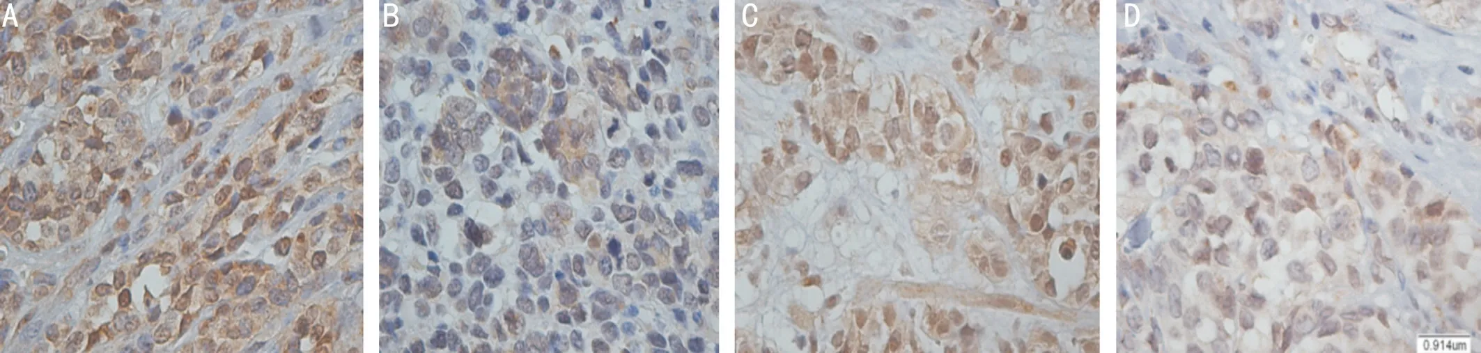
HPSE-1 protein expression was examined in 28 orbital ERMS and 23 orbital ARMS by immunohistochemistry. Positive expression was observed with different staining intensity in 92.9% of embryonal samples and 91.3% of alveolar samples tested and primarily located in the cytoplasm and nucleus. Generally speaking, there was no significant difference in HPSE‐1 immunostaining intensity between ERMS and ARMS specimens. However, both of them displayed the similar tendency. Tissue from untreated(no chemoradiotherapy before surgery) RMS patients tended to show intense staining, whereas tissue from RMS patients treated with chemoradiotherapy (chemoradiotherapy followed by surgery) showed relatively weaker staining (Figure 2).
Current knowledge about HPSE-1 mRNA and protein expression in orbital ARMS is only based on our previous report. However, it has not been elucidated whether HPSE-1 protein can be a targeted therapy factor of orbital RMS. In the present study, we examined the expression of HPSE-1 protein levels in ERMS and ARMS specimens, as well as preoperative chemoradiotherapy and untreated patients. Furthermore, the antitumor efficacy of HPSE‐1 targeted therapy was investigated. Both RMS cells viability and tube formation of human umbilical vein endothelial cells (HUVECs) induced by vascular endothelial growth factor (VEGF) were evaluated.
RESULTS
Basement membrane extracellular matrix (Matrigel; BD Biosciences, San Jose, CA, USA) was thawed at 4°C overnight. The 200 μL pipette tips and a 96‐well plate were also kept at 4°C overnight. Matrigel (90 μL) was loaded in each well, and then the plate was incubated at 37°C for 30min to solidify the matrix. The appropriate amount of recombinant lentivirus containing HPSE-1 shRNA (1×10TU/mL)and negative control lentivirus (1×10TU/mL) were added into the cells (MOⅠ=30), and the final concentration of polybrene was 8 μg/mL. After infection 48h, the pretreated HUVECs were seeded on the solidified matrigel at a density of 10cells/well.The plates were kept at room temperature for 15min and then transferred to the 5% COincubator at 37°C. The capillary-like structure formation of HUVECs was observed at different time points during a 12h experimental period using a microscope.After incubation for 4, 6, and 10h, the capillary-like tube formation was quantified by counting the length of tubes in 5 randomly selected optical fields using an Olympus microscope(Olympus Corporation, Tokyo, Japan).
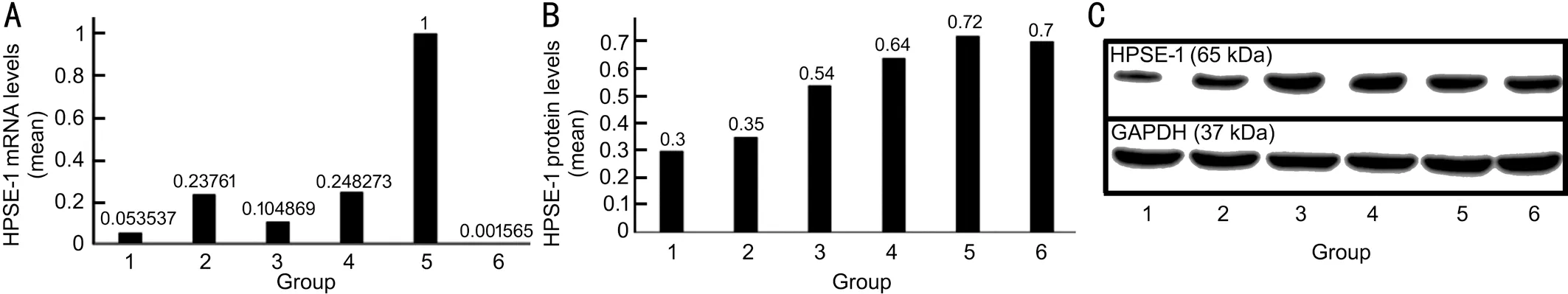
Four recombinant plasmids of HPSE-1 shRNA (1-4), targeting four different encoding regions of HPSE-1 mRNA, were successfully constructed using pcDNA6.2-GW/EmGFP-miR,which were confirmed by DNA sequence analysis. Forty‐eight hours after the co-transfection of HPSE-1 shRNA plasmid and HPSE-1 eukaryotic expression vector, 293T cells were divided into six groups,., HPSE-1 shRNA1-4, negative control,and blank cells group. All the four recombinant plasmids of HPSE-1 shRNA successfully down-regulated HPSE-1 mRNA and protein expression levels, and the plasmid with HPSE-1 shRNA1 was shown to be the most efficient RNAi vector.Compared with negative control group, HPSE-1 mRNA and protein expression levels in 293T cells that were transfected with HPSE-1 shRNA1 were down-regulated by maximum 95% (RT-qPCR) and 58.3% (Western blotting) respectively(Figure 3).
以石英為主的礦床主要有熱液石英脈型礦床、偉晶花崗巖型礦床、沉積硅質砂巖型礦床以及變質沉積石英巖礦床,這類礦床經開采選別后所產生的尾礦中SiO2的含量一般為60%~90%。常見如金礦和鐵礦。石英脈型金礦是我國主要的金礦工業類型,其數量和儲量分別占金礦床總數量和金總儲量的50%以上[5]。我國最大的高硅鞍山型鐵礦,其尾礦中SiO2含量可達83%,如本鋼、鞍鋼、首鋼、太鋼和唐鋼等公司下屬選礦廠排出的尾礦;據資料顯示,鞍鋼礦山公司尾礦庫尾礦中的SiO2含量在80%左右[6-8]。
2.健全強制免疫申報機制,推行 “主動免疫”。自2007年以來,南川區推行兩種免疫模式,一是全面推行以農村散養戶為主的“仔豬閹割三針二次強化免疫”的程序,即要求畜主在母豬產仔后提前主動到鄉鎮畜牧獸醫站申報登記,站上及時安排包片獸醫按閹割免疫程序服務好;二是……
登錄APP查看全文
 International Journal of Ophthalmology
2022年1期
International Journal of Ophthalmology
2022年1期
- International Journal of Ophthalmology的其它文章
- lnstructions for Authors
- Comment on: Trends in research related to high myopia from 2010 to 2019: a bibliometric and knowledge mapping analysis
- Progress of clinical therapies for dry age-related macular degeneration
- Observation seasonal variation of intraocular pressure in young healthy volunteers
- Effectiveness of oral probiotics supplementation in the treatment of adult small chalazion
- Characterization and validation of a chronic retinal neovascularization rabbit model by evaluating the efficacy of anti-angiogenic and anti-inflammatory drugs