Effect of miR-184 and miR-205 on the tumorigenesis of conjunctival mucosa associated lymphoid tissue lymphoma through regulating RasL10B and TNFAlP8
2022-01-20 07:03:46YuZhenLiPeiMouYaShenLianDiGaoXinXinChenRuiLiWei
INTRODUCTION
Five samples of tumor tissue from patients (4 males and 1 female, mean age 60.6±12.2y, range 40 to 71y, the onset time of 3mo to 6y) with conjunctival MALT lymphoma in this study were collected in Shanghai Changzheng Hospital from July to October in 2016, during anterior open acquisition orbital tumor surgery for MALT lymphoma. The matched adjacent normal conjunctival tissues as controls were obtained and were snap-frozen for further analysis. All patients had no other diseases except MALT lymphoma.
RPMI8226 cells were planted 1.0×10cells per well in 6‐well plates. When culturing to 50% confluence,miR-184 and miR-205 mimics or their negative control were transfected into cells carried by Lipofectamine 2000,respectively. Cell apoptosis was examined by the Annexin V-FITC/PI apoptosis detection kit, and detected by a flow cytometer and Cell Quest Pro version 6.0 software (FACScan;BD Biosciences, Franklin Lakes, NJ, USA).
MATERIALS AND METHODS
miR-184 is involved in regulating the tumorigenesis and metastasis of tumor, as a new member of miRNA family.Previous research showed that the expression level of miR-184 was decreased in various types of tumor. In glioma tumor tissues, the expression of miR‐184 was significantly decreased compared with normal brain tissue. In addition, Liangfound that the level of miR-184 was reduced in central nervous system lymphoma. Meanwhile, the expression of miR-184 was found increased in other types of tumor. miR-184 was extensively expressed in PANC-1 cells, as a pancreatic ductal adenocarcinoma (PDAC) cell line.
由于生態(tài)和區(qū)位的不同,北京城市副中心相對(duì)應(yīng)的每個(gè)鎮(zhèn)變化的建設(shè)用地均不相同,其變化的建設(shè)用地面積大小代表了2030年期限內(nèi)每個(gè)鎮(zhèn)的發(fā)展?jié)摿ΑR虼耍鞘性鲩L(zhǎng)模擬的各鎮(zhèn)發(fā)展?jié)摿A(chǔ)可作為考慮各鎮(zhèn)區(qū)的主觀發(fā)展需求的依據(jù),在此基礎(chǔ)上可反推出未來(lái)通州各鎮(zhèn)集體建設(shè)用地的減量面積,作為未來(lái)通州各鎮(zhèn)集體建設(shè)用地指標(biāo)分配的參考條件。規(guī)劃中,將北京城市副中心各鎮(zhèn)的建設(shè)用地變化量進(jìn)行計(jì)算,得出各鎮(zhèn)的現(xiàn)狀建設(shè)用地變化比例。集體建設(shè)用地指標(biāo)轉(zhuǎn)換流程如圖4所示。
Ocular adnexal lymphoma (OAL) is the most common tumors in the eyes. Extranodal marginal zone B-cell lymphoma of mucosa-associated lymphoid tissue (MALT), as a rare form of non-Hodgkin’s lymphoma, is the majority subtype of OAL. The OAL causes a serious threaten to patient health.Therefore, the development of novel target drug is important for patients with OALs. However, the pathological mechanism of OALs is not clearly. MicroRNAs (miRNAs), as a group of short non-coding RNA molecules with 21-25 nucleotides length, regulate target gene translation or degradation through interacting with complementary sites in the 3’ untranslated region (3’UTR) of target mRNAs. Increasing evidences indicated that miRNAs were involved in the pathogenesis of MALT lymphoma. miR-142 and miR-155 regulate the MALT pathogenesis through repressing the target gene TP53INP1. The decreased expression of miR-34a induces the target genes increase, such as FOXP1, p53, and BCL2, to regulate MALT lymphoma development and apoptosis. miR-200 is up-regulated and suppressed the target protein cyclin E2 in conjunctival MALT lymphoma.
RPMI8226 cells were transfected with miR-205 mimics, miR-205 inhibitor or negative control miRNA using INTERFER in transfection reagent following the manufacturer’s instructions (Polyplus). Whole cell extracts for immunoblotting analysis were prepared as previously reported.These antibodies were used for immunoblotting studies:rabbit anti RASL10B (1:500, Abbkine), rabbit anti TNFAIP8(1:1000, Sigma), rabbit anti GAPDH (1:10 000, Sigma), goat anti rabbit IgG-HRP (1:6000, Millipore).
Previous studies indicated that miR-205 and miR-184 were involved in tumor proliferation, apoptosis, invasion, and metastasis. miR-205 acts as tumor activator or suppressor in various types of tumortargeting various genes. The increased miR-205 expression promotes the proliferation,invasion, and migration of nasopharyngeal carcinoma cells.In renal cell carcinoma cells, miR-205 expression is decreased.In renal cell carcinoma cells, PTEN expression is upregulated and p-AKT expression is downregulated after the miR-205 mimics transfection. Previous studies indicated that miR-184 inhibits tumor cells proliferation and invasion in glioma and non-small cell lung cancer cells. Our previous study indicated that the expressions of miR-205 and miR-184 were decreased greatly in MALT tissue compared with control tissue. However, the mechanism of miR-205 and miR-184 regulating MALT lymphoma was not clear.
Transfected cells were gathered and then suspended. For the next step, 5×10or 1×10of the cells were planted onto the upper part of Transwell chambers (Corning, NY, USA), along with or without Matrigel covering (BD Biosciences, SanDiego, CA, USA). Ten percent of FBS was added into the media and placed into the underlying part as a chemo attractant. Twelve or twenty-four hours later, cells migrated or invaded into the underlying part were recorded by an inverted microscope (Olympus, Tokyo, Japan).
患者均展開(kāi)腹彩超多普勒超聲的檢查,同時(shí)進(jìn)行診斷結(jié)果、手術(shù)病理證實(shí)的比較,選擇GE型LOGIQP5與多普勒超聲診斷儀進(jìn)行,對(duì)探頭頻率進(jìn)行選擇時(shí),需要按照2~9MHz標(biāo)準(zhǔn)進(jìn)行[2]。患者以平臥位、側(cè)臥位狀態(tài),保證膀胱充盈。選擇彩色多普勒超聲探頭置于患者腹壁,在對(duì)患者胎兒和羊水進(jìn)行了解后,對(duì)胎盤(pán)邊緣、子宮頸間關(guān)系進(jìn)行了解。在對(duì)探頭方向進(jìn)行相應(yīng)調(diào)整后,引導(dǎo)患者正確進(jìn)行體位變化,仔細(xì)觀察胎盤(pán)邊緣和子宮頸內(nèi)關(guān)系,以便于進(jìn)行有效診斷與分類。最后進(jìn)行胎盤(pán)間隙與胎盤(pán)實(shí)質(zhì)、周邊血流的觀察,同時(shí)掌握胎盤(pán)植入情況[3]。
Total RNA was collected by TRIzol reagent (Invitrogen). M-MLV reverse transcriptase (RT; Promega, Madison, WI, USA) was then used to reverse-transcribe. RT primers for miR-205 or random primers (Promega) for TNFAIP8 were from RiboBio(Guangzhou, China). Bio-Rad CFX96 Touch sequence detection system (Bio-Rad Laboratories Inc, Hercules, CA,USA) was used to conduct the quantitative polymerase chain reaction (qPCR) reactions. Platinum SYBRGreen qPCR SuperMix-UDG reagents (Invitrogen) were added into the reaction. Experiments were done three times. U6 or GAPDH were setting as controls. Primers used for qPCR were as followed: Urp-re-R2: 5’-GTGCAGGGTCCGAGGT-3’,miR182-loop-2: 5’-GTCGTATCCAGTGCAGGGTCC GAGGTATTCGCACTGGATACGACAGAGTGTG-3’,miR182-re-F-2: 5’-CGGCGTTTGGCAATGGTAGAAC-3’,miR205-loop-2: 5’-GTCGTATCCAGTGCAGGGTCCGA GGTATTCGCACTGGATACGACCACAGAC-3’, miR205-re-F-2: 5’-CGGCGTCCTTCATTCCACCG-3’, RasL10B-F:5’-GGGGGTACCATGGTCTCCACC-3’, RasL10B-R:5’-GCGGGATCCGCTCGGCCAG-3’, TNFAIP8-F:5’-TTCCATCAGGTGGATTATAC-3’, TNFAIP8-R:5’-AGGTGGCGCTGAATGATTTG-3’.
Human B cell line RPMI8226 (ATCC,Rockville, MD, USA) were cultured in RPMI 1640 medium supplemented with 10% fetal bovine serum (FBS) and maintained at 37℃ in a humidified atmosphere containing 5%CO. The HEK cell line is a commonly used model cell which has a relatively high transfection rate, while the RPMI8226 cell line, a commonly used human B cell line, is utilized to verify the effect of miRNAs on B cells.
The psiCHECK-2 luciferase reporter plasmid (Promega) carried the cloning RASL10B WT and Mt 3’UTR , TNFAIP8 WT and Mt 3’UTR. Cells were first planted into six‐well plates. One day later, cells were co‐transfected using Lipofectamine 2000 reagent (Invitrogen),with the following: RASL10B WT or Mt 3’UTR reporter plasmids and miR-184 mimic, TNFAIP8 WT or Mt 3’UTR reporter plasmids and miR-205 mimic, and also the control vector pRL-TK (Promega). For the activities of luciferase,results were measured using the Dual-Luciferase Reporter Assay System (Promega). The primers used in luciferase assay as followed: Rasl10b WT-F: 5’-TCGAGCAGCCTTAACTCG ATGGTCCGTCCCTGCCAGGTGCCGC-3’, Rasl10b WT-R:5’-GGCCGCGGCACCTGGCAGGGACGGACCATCGAGT TAAGGCTGC-3’; Rasl10b Mut-F: 5’-TCGAGCAGCCTTA ACTCGATGGAGGCAGCCTGCCAGGTGCGC-3’, Rasl10b Mut-R: 5’-GGCCGCGCACCTGGCAGGCTGCCTCCATCG AGTTAAGGCTGC-3’; TNFAIP8 WT-F: 5’-TCGAGAAGA TGGAGCACTGCTGATTTATGAAGGAAAAAAGAGC-3’,TNFAIP8 WT-R: 5’-GGCCGCTCTTTTTTCCTTCATAAAT CAGCAGTGCTCCATCTTC-3’; TNFAIP8 Mut-F: 5’-TCG AGAAGATGGAGCACTGCTGATTTTACTTCGAAAAAA GAGC-3’, TNFAIP8 Mut-R: 5’-GGCCGCTCTTTTTTCGA AGTAAAATCAGCAGTGCTCCATCTTC-3’; Rasl10b WTF: 5’-CAGCCUUAACUCGAUGGUCCGUCCCUGCCAG GUGCC-3’, Rasl10b Mut: 5’-CAGCCUUAACUCGAUGG AGGCAGCCUGCCAGGUGC-3’; TNFAIP8-WT: 5’-AAGA UGGAGCACUGCUGAUUUAUGAAGGAAAAAAGA-3’,TNFAIP8-Mut: 5’-AAGAUGGAGCACUGCUGAUUUUAC UUCGAAAAAAGA-3’.
All statistical analysis were completed by the SPSS 15.0 statistical software (SPSS Inc., Chicago,IL, USA). Difference of measurement data was compared with-test or one-way ANOVA.<0.05 were considered statistically significant.
RESULTS
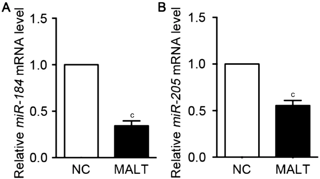
RNA was extracted from tumor tissues and adjacent tissues of 5 patients with MALT, and the mRNA expression of miR-184 and miR-205 was detected by qRT-PCR. The results showed that the expression of miR-184 and miR-205 mRNA were significantly downregulated in the lymphoma tissues compared with the adjacent tissues (<0.001; Figure 1).
Ⅰn order to study the effect of miR-184 and miR-205 on the proliferation of lymphoma cells,miR-184 mimics and corresponding negative controls were transfected into the RPMI8226 cells. The CCK8 kits were used after transfection of 24, 48, 72, and 96h, and the value with 450 nm wavelength was detected. The results showed that the transfection of miR184 mimics notably inhibited the proliferation rate of RPMI8226 cells than the negative group.A similar result was observed when miR-205 mimics was transfected into the RPMI8226 cells. The results showed that the proliferation rate of RPMI8226 cells was significantly inhibited after transfected with miR-205 mimics than with the negative group. These results indicate that miR-184 and miR-205 notably inhibit the proliferation of RPMI8226 cells.
Then, the effect of miR-184 and miR-205 on the apoptosis of lymphoma cells was explored by flow cytometry. The results showed that the apoptosis rate was 0.06%±0.006% in normal RPMI8226 cells group, and the rate was 1.2%±0.73%in negative control group. Apoptosis rate was 12.4%±0.32%in miR-184 mimics treated group, and the rate of the miR-205 mimics treated group was 17.1%±0.42%. Compared with normal group and negative control group, miR-184 and miR-205 mimics induced the apoptosis rate of RMPI8226 cells increased significantly (Figure 2;<0.001).
Previous study has shown that RasL10B may be a downstream target protein of miR-184 which participates in the pathogenesis of MALT lymphoma.We detected the expression level of RasL10B in MALT lymphoma tissues by real-time qPCR and Western blotting.The results revealed that RasL10B mRNA and protein in MALT lymphoma were significantly decreased compared para-cancerous tissue (Figure 5A-5C). The results of luciferase test showed that after transfection of miR184 mimics, the luciferase expression was significantly reduced (<0.01) after transfection of the wild type RasL10B gene 3’UTR compared with the control group and the mutant group, indicating that miR184 has direct regulation on 3’UTR of the RasL10B gene(Figure 5D). In terms of the effect on the control group, the protein expression level of RasL10B was raised in RPMI8226 cells transfected to miR-184 mimics. However, miR184 inhibitor obviously decreased the expression level of RasL10B protein (<0.05; Figure 5E, 5F). These results suggested that miR184 can promote the gene expression of RasL10B in MALT lymphoma and RPMI8226 cells.
Furthermore, we detected the effect of miR-184 and miR-205 on the invasion of RPMI8226 cells. The results showed that comparison to the negative group, the number of invaded cells in the miR-184 inhibitor treated groups was 142.2%±21.3%, while the number in mimics treated group was only 68.1%±12.7% (Figure 4A, 4C). After transfection of 72h, compared to the negative control group, the number of invaded cells in the miR-205 mimics treated group was only 59.2%±12%, while the number of miR205 inhibitor groups through the cells was 143.4%±24.1% in the control group(Figure 4B, 4D). These results suggested that miR-184 and miR-205 attenuated the ability of migration and invasion of RPMI8226 cells.
The effect of miR-184 and miR-205 on migration of RPMI8226 cells were examined by Transwell assay. The results showed that comparison to the negative group (100%), the number of migrated cells was the 118%±4.9% in the miR-184 inhibitor treated group. However,the number of migrated cells only was 61.4%±12.7% in the miR-184 mimics treated group (Figure 3A, 3C). Comparison to the negative control group (100%), the number of migrated cells in miR-205 inhibitor treated group was 110.7%±4.5%,while the number of migrated cells in the miR-205 mimics treated group was only 53%±11.7% (Figure 3B, 3D).
選擇葡萄酒的人有千萬(wàn)種因由,但這一路馳騁,從未動(dòng)搖或懈怠的人真的是鳳毛麟角,連我,也曾一度因?yàn)榭床磺逑乱粋€(gè)目標(biāo)而出走過(guò),才意識(shí)到我們根本沒(méi)有完整地看清這個(gè)行業(yè)的路徑,目標(biāo)也定得太過(guò)短小,但他的路徑卻一早就查好了。……
登錄APP查看全文
猜你喜歡
保健醫(yī)苑(2021年7期)2021-08-13 08:48:02
學(xué)生天地(2020年36期)2020-06-09 03:12:30
小學(xué)科學(xué)(學(xué)生版)(2020年5期)2020-05-25 07:11:32
小學(xué)科學(xué)(學(xué)生版)(2020年4期)2020-05-21 07:30:46
小學(xué)科學(xué)(學(xué)生版)(2020年3期)2020-03-25 13:31:22
中國(guó)外匯(2019年18期)2019-11-25 01:41:56
電子制作(2018年14期)2018-08-21 01:38:28
人大建設(shè)(2017年10期)2018-01-23 03:10:17
民生周刊(2017年19期)2017-10-25 10:29:03
領(lǐng)導(dǎo)文萃(2015年4期)2015-02-28 09:19:05
 International Journal of Ophthalmology
2022年1期
International Journal of Ophthalmology
2022年1期
- International Journal of Ophthalmology的其它文章
- lnstructions for Authors
- Comment on: Trends in research related to high myopia from 2010 to 2019: a bibliometric and knowledge mapping analysis
- Progress of clinical therapies for dry age-related macular degeneration
- Observation seasonal variation of intraocular pressure in young healthy volunteers
- Effectiveness of oral probiotics supplementation in the treatment of adult small chalazion
- Characterization and validation of a chronic retinal neovascularization rabbit model by evaluating the efficacy of anti-angiogenic and anti-inflammatory drugs