siRNA干擾ΔNp63基因表達對人宮頸癌Siha細胞增殖及凋亡影響研究
2017-01-18 09:03:23周福興劉海霞羅天愛陳必良
中國婦幼健康研究 2016年12期
關鍵詞:檢測
李 娜,劉 玉,馬 芮,周福興,劉海霞,羅天愛,陳必良
(第四軍醫大學西京醫院婦產科,陜西 西安 710032)
?
siRNA干擾ΔNp63基因表達對人宮頸癌Siha細胞增殖及凋亡影響研究
李 娜,劉 玉,馬 芮,周福興,劉海霞,羅天愛,陳必良
(第四軍醫大學西京醫院婦產科,陜西 西安 710032)
目的 通過抑制宮頸癌細胞株Siha中ΔNp63的表達,探討ΔNp63對宮頸癌細胞增殖及凋亡的影響。方法 將宮頸癌細胞株Siha分為實驗組和對照組,實驗組轉染ΔNp63的特異性siRNA,對照組轉染陰性對照siRNA。實時熒光定量PCR(QRT-PCR)及蛋白質印跡法檢測Siha細胞轉染前后ΔNp63基因在mRNA及蛋白水平的變化;CCK8法檢測細胞增殖,流式細胞儀測定細胞凋亡率。結果 實驗組Siha細胞轉染siRNA后,ΔNp63的mRNA表達為0.32±0.06,低于陰性對照組0.95±0.08(t=10.92,P<0.05)。實驗組Siha細胞轉染siRNA后,ΔNp63的蛋白表達為0.32±0.09,低于陰性對照組1.00±0.06(t=9.78,P<0.05)。實驗組Siha細胞生長速度明顯低于陰性對照組,實驗組Siha細胞凋亡率為2.13±0.75,低于陰性對照組14.19±1.36(t=15.36,P<0.05)。結論 人宮頸癌細胞株中存在ΔNp63的表達,特異性siRNA可下調宮頸癌細胞株Siha中ΔNp63基因的表達,對細胞的增殖具有負性調節作用,并能誘導細胞凋亡。
宮頸癌;ΔNp63;RNA干擾;細胞增殖;細胞凋亡
p63是近年發現的p53 家族成員之一。由于啟動子的不同和3'端剪切方式的不同,p63基因編碼多種具有不同活性的異構體,可分成兩大類:具有反式激活區的TA異構體和N 末端截短的ΔN異構體[1]。TA 異構體具有p53 樣活性誘導細胞周期停滯和凋亡,ΔN異構體可以抑制p53的功能促進轉化細胞的生長[2-3]。最初發現,ΔNp63是鱗狀上皮基底細胞中極為重要的表達蛋白,可拮抗p53的抑瘤作用,在許多的上皮惡性腫瘤中都有過表達。目前,有關ΔNp63在宮頸癌中的作用機制報道尚少。為了研究ΔNp63在宮頸癌中發揮的作用,本研究利用RNA干擾(RNA interference,RNAi)技術,針對人ΔNp63基因設計并合成小干擾RNA用以沉默ΔNp63基因,觀察ΔNp63基因對宮頸癌細胞Siha細胞增殖和凋亡能力的影響。
1 資料與方法
1.1材料
宮頸癌細胞株Siha細胞為第四軍醫大學西京醫院婦產科實驗室保存,小鼠抗人ΔNp63單克隆抗體、小鼠抗人甘油醛-3-磷酸脫氫酶(glyceraldehyde-3-phosphate dehydrogenase,GAPDH)單克隆抗體購自BioLegend公司,極限最低培養基(minimum essential medium,MEM)培養基購自HyClone公司,胎牛血清購自浙江天杭公司,脂質體Lipo-2000購自Invitrogen公司,細胞計數試劑盒(Cell Counting Kit-8,CCK8)購自上海七海復泰生物公司,逆轉錄試劑盒、實時定量-聚合酶鏈反應(real-time polymerase chain rection,RT-PCR)試劑盒購自大連TaKaRa公司。ΔNp63引物序列:上游5′-CCAAAGCGAGGCACCCTTAC-3′;下游5′-CATTGAGCTG ̄AGGCCACAAGA-3′。內參Actin引物序列:5′-ACAGAGC ̄CTCGCCTTTGC-3′;下游5′-CGCGGCGATATCATCATCCA-3′。 針對ΔNp63的特異性siRNA序列為5′-UGCCCAGAC ̄UCAAUUUAGU-3′[4],陰性對照siRNA序列為5′-UUCUC ̄CGAACGUGUCACGUTT-3′。引物及siRNA均委托上海生工公司合成。流式細胞術檢測在第四軍醫大學口腔醫院實驗室進行。
1.2細胞培養
Siha細胞培養于含10%胎牛血清的MEM培養基中,在37°、5% CO2的培養箱中常規培養,并取對數生長期的細胞用于實驗。
當宮頸癌Siha細胞生長密度為80%~90%時,胰酶消化后均勻鋪于6、96孔板上,按照Lip-2000試劑說明書將特異性siRNA及對照組siRNA分別轉染到Siha細胞中,其轉染終濃度均為100nmol/L。
1.3 siRNA瞬時轉染
取對數生長期的Siha細胞按每孔1×10⁵的密度接種于6孔板中,待細胞生長至60%時進行轉染。將Siha細胞分為兩組:實驗組和對照組,分別轉染特異性siRNA和陰性對照siRNA。轉染時用Opti-MEM無血清培養基稀釋Lip-2000與siRNA序列,具體按Lip-2000說明書進行,轉染終體積為每孔2mL培養基,siRNA轉染終濃度為每孔100nmol/L。轉染6小時后換正常培養基,轉染48h后,收集細胞,進行相應實驗。
1.4實時定量PCR檢測
按1.3方法轉染48h后,按照TaKaRa RNA提取試劑盒提取Siha細胞的總RNA,細胞RNA的提取嚴格按照說明書在冰上進行,以防RNA降解。取A260/280值在1.8~2.0之間的純度較好的RNA原液放置于-80°保存。測量RNA濃度,逆轉錄為cDNA,進行RT-PCR反應。反應體系為20μL體系,反應條件:95° 3min,95°變性30s,55°退火20s,72°延伸20s ,40個循環,最后72°再次延伸10min。實驗重復3次。
1.5蛋白印跡法檢測
按1.3方法轉染48h后,取實驗組及對照組細胞置于冰上裂解細胞提取總蛋白并進行BCA定量,調整蛋白濃度。按每孔30μg總蛋白量上樣,行SDS-聚丙烯酰胺凝膠電泳,300mA電流恒轉2h,5%脫脂奶粉封閉2h,抗ΔNp63和抗GAPDH抗體 4°孵育過夜,次日用相應二抗室溫孵育2h,滴加化學發光底物進行檢測,并用Quantity One軟件進行分析。實驗重復3次。
1.6細胞增殖檢測
采用CCK8方法進行細胞增殖實驗。取對數生長期的Siha細胞按每孔103的密度接種于96孔板中,每孔取5個平行孔進行檢測,培養過夜后,按1.3的轉染方法進行轉染,分組同1.3。轉染48h后每孔按100:10比例加入正常培養基和CCK8試劑,37°孵育2h,在酶聯免疫檢測儀上450nm下測各孔的吸光度值(optical density,OD450)。對轉染6h、24h、48h、72h分別進行檢測。實驗重復3次。
1.7細胞凋亡檢測
采用流式細胞儀檢測細胞凋亡。取對數生長期Siha細胞按每孔1×10⁵的密度接種于6孔板中,轉染方法及分組同1.3。待轉染48h后,用不含乙二胺四乙酸(ethylene dDiamine tetraacetic acid,EDTA)的胰酶消化后離心,收集細胞。用冷PBS洗滌細胞兩次,盡可能吸盡PBS。用400μL l×Annexin V結合液懸浮細胞,濃度大約為1×10⁶。在細胞懸液中加入5μL Annexin V-FITC染色液,輕輕混勻于2~8°避光條件下孵育15min。加入10μL PI染色液輕輕混勻后2~8°避光條件下孵育5min。立即用流式細胞儀檢測。實驗重復3次。
1.8 統計學方法

2結果
2.1特異性siRNA對ΔNp63 mRNA表達水平的影響
特異性siRNA轉染48h后,RT-PCR檢測結果顯示,實驗組和對照組ΔNp63 mRNA的表達分別為0.32±0.06和0.95±0.08,兩組相比差異有統計學意義(t=10.92,P<0.05),見圖1。

圖1 RT-PCR方法檢測特異性siRNA對Siha細胞ΔNp63 mRNA表達的影響
Fig.1 RT-PCR analysis of the effect of siRNA on ΔNp63 mRNA expression in Siha cells
2.2特異性siRNA對ΔNp63蛋白表達水平的影響
特異性siRNA轉染48h后,Western blot檢測結果顯示,實驗組和對照組ΔNp63蛋白表達水平分別為0.39±0.09和1.00±0.06,兩組相比差異有統計學意義(t=9.78,P<0.05),見圖2。

圖2 Western blot方法檢測特異性siRNA對Siha細胞ΔNp63蛋白表達的影響
Fig.2 Western blot analysis of the effect of siRNA on ΔNP63 protein expression in Siha cells
2.3特異性siRNA干擾ΔNp63表達對細胞增殖的影響
用CCK8法分別檢測轉染6h、24h、48h、72h后Siha細胞的增殖情況,檢測結果顯示,實驗組和對照組OD490/6h分別為0.23±0.04、0.26±0.02;OD490/24h分別為0.39±0.04、0.52±0.06;OD490/48h分別為0.74±0.04、1.04±0.05;OD490/72h分別為0.99±0.03、1.52±0.03。在轉染24h、48h、72h后,所測吸光度值有統計學意義。實驗組細胞生長曲線較為平緩,細胞生長速度較對照組相比明顯減慢(t6h=1.16,P>0.05;t24h=3.12,P<0.05;t48h=8.12,P<0.05;t72h=21.64,P<0.05),見圖3。
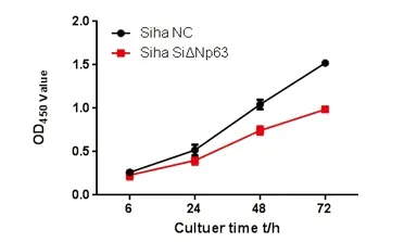
圖3 CCK8法檢測特異性siRNA干擾ΔNp63 基因后Siha細胞的生長曲線
Fig.3 CCK8 analysis of growth curve of Siha cells after siRNA interfering ΔNp63 gene
2.4特異性siRNA干擾ΔNp63表達對細胞凋亡的影響
特異性siRNA轉染48h后進行流式細胞儀檢測實驗,結果顯示,實驗組和對照組Siha細胞凋亡率分別為14.19±1.36和2.13±0.75,t=15.36,P<0.05,差異有統計學意義,見圖4。

圖4 流式細胞儀檢測特異性siRNA對Siha細胞凋亡的影響
Fig.4 FACS analysis of the effect of siRNA on Siha cells apoptosis
3討論
3.1 p63基因的結構與功能
p63分為TA和ΔN兩個亞型,根據C端的不同剪切TA和ΔN又分為α、β、γ三種亞型[5-7]。ΔNp63與p53的同源性高達60%,如核心DNA結合區、寡聚化區,與p53不同的是ΔNp63還具有獨特的N-末端的轉錄激活區和C-末端SAM區[8-9]。有研究表明,ΔNp63可能是通過與p53競爭性結合DNA靶點區來拮抗p53的抑癌作用,從而通過對增殖、凋亡進行調節而影響腫瘤的發生發展[9]。因此,ΔNp63蛋白并不是一種腫瘤抑制基因,而是致癌基因。p63主要分布于各種上皮組織的增生區,如食管、口腔黏膜、皮膚、子宮頸的復層膜和生精小管上皮都有很強的核染色,其中轉錄活性最強的是ΔNp63[10]。近年來,越來越多的學者發現,ΔNp63的過表達與多種腫瘤的發生相關,如口腔癌、頭頸部癌、肺癌、膀胱癌等[11-14]。在宮頸癌組織中,幾乎所有的宮頸癌細胞都表達ΔNp63蛋白,提示ΔNp63蛋白與宮頸癌的發生發展密切相關[15]。
3.2利用RNAi技術下調ΔNp63的表達后,對宮頸癌細胞生物學功能的影響
本研究通過RNAi技術,將特異性的siRNA轉染入人宮頸癌細胞株Siha中,通過RT-PCR技術和蛋白質印跡技術分別檢測特異性siRNA干擾后ΔNp63 mRNA和蛋白的表達情況。結果顯示,轉染siRNA后,Siha細胞的ΔNp63 mRNA、蛋白的表達水平均明顯降低,提示特異性siRNA可有效抑制Siha細胞中ΔNp63基因的表達。隨后,我們利用RNAi技術對ΔNp63在細胞增殖及凋亡中的作用進行了初步探討,結果顯示轉染特異性siRNA的實驗組與轉染陰性對照siRNA的對照組相比,其增殖率明顯受到抑制,凋亡率顯著增加。
本研究結果表明,宮頸癌細胞中存在ΔNp63的表達,特異性siRNA可有效抑制ΔNp63基因的表達,并抑制細胞增殖,促進細胞凋亡,但其機制目前并不明確。本研究為進一步的機制研究提供了一定的實驗基礎。
[1]Bornachea O,López-Calderón F F,Dueas M,etal.The downregulation of ΔNp63 in p53-deficient mouse epidermal tumors favors metastatic behavior[J].Oncotarget,2015,6(27):24230-24245.
[2]Zhao W,Wang H,Han X,etal.ΔNp63α attenuates tumor aggressiveness by suppressing miR-205/ZEB1-mediated epithelial-mesenchymal transition in cervical squamous cell carcinoma[J]. Tumour Biol,2016,37(8):10621-10632.
[3]Arason A J,Jonsdottir H R,Halldorsson S,etal.deltaNp63 has a role in maintaining epithelial integrity in airway epithelium[J].PLoS One,2014.9(2):e88683.
[4]Suarez-Carmona M,Hubert P,Gonzalez A,etal.ΔNp63 isoform-mediated β-defensin family up-regulation is associated with (lymph)angiogenesis and poor prognosis in patients with squamous cell carcinoma[J].Oncotarget,2014,5(7):1856-1868.
[5]Bid H K,Roberts R D,Cam M,etal.ΔNp63 promotes pediatric neuroblastoma and osteosarcoma by regulating tumor angiogenesis[J].Cancer Res,2014,74(1):320-329.
[6]Moergel M,Goldschmitt J,Stockinger M,etal.ΔNp63 expression in four carcinoma cell lines and the effect on radioresistance--a siRNA knockdown model[J].Clin Oral Investig,2014,18(4):1259-1268.
[7]Kim K H,Cho E G,Yu S J,etal.ΔNp63 intronic miR-944 is implicated in the ΔNp63-mediated induction of epidermal differentiation[J].Nucleic Acids Res,2015,43(15):7462-7479.
[8]Ho J Y,Chang F W,Huang F S ,etal.Estrogen Enhances the Cell Viability and Motility of Breast Cancer Cells through the ERα-ΔNp63-Integrin β4 Signaling Pathway[J].PLoS One,2016,11(2):e0148301.
[9]Lena A M,Duca S,Novelli F,etal.Amino-terminal residues of ΔNp63, mutated in ectodermal dysplasia, are required for its transcriptional activity[J].Biochem Biophys Res Commun,2015,467(2):434-440.
[10]Orzol P,Nekulova M,Holcakova J,etal.ΔNp63 regulates cell proliferation, differentiation, adhesion, and migration in the BL2 subtype of basal-like breast cancer[J].Tumour Biol,2016,37(8):10133-10140.
[11]Curtis K M,Aenlle K K,Frisch R N,etal.TAp63γ and ΔNp63β promote osteoblastic differentiation of human mesenchymal stem cells: regulation by vitamin D3 metabolites[J].PLoS One,2015,10(4):e0123642.
[12]Kakuki T,Kurose M,Takano K,etal.Dysregulation of junctional adhesion molecule-A via p63/GATA-3 in head and neck squamous cell carcinoma[J].Oncotarget,2016,7(23):33887-33900.
[13]Bretz A C,Gittler M P,Charles J P,etal.ΔNp63 activates the Fanconi anemia DNA repair pathway and limits the efficacy of cisplatin treatment in squamous cell carcinoma[J].Nucleic Acids Res,2016,44(7):3204-3218.
[14]Gaya J M,López-Martínez J M,Karni-Schmidt O,etal.ΔNp63 expression is a protective factor of progression in clinical high grade T1 bladder cancer[J].J Urol,2015,193(4):1144-1150.
[15]Zhou Y,Xu Q,Ling B,etal.Reduced expression of ΔΝp63α in cervical squamous cell carcinoma[J].Clin Invest Med,2011,34(3):E184-E191.
[專業責任編輯:楊筱鳳]
Effects of small RNA interfering the expression of ΔNp63 on the proliferation and apoptosis in Siha cells
LI Na, LIU Yu, MA Rui, ZHOU Fu-xing, LIU Hai-xia, LUO Tian-ai, CHEN Bi-liang
(Department of Gynecology and Obstetrics, Xijing Hospital, Fourth Medical University, Shaanxi Xi’an 710032, China)
Objective To detect the effect of ΔNp63 on proliferation and apoptosis on human cervical cancer cells by suppressing the expression of ΔNp63 in cell line Siha. Methods Siha cell lines were divided into experimental group (siRNA with transfected ΔNp63) and control group (transfected negative controlled siRNA). Real-time reverse transcription-polymerase chain reaction (RT-PCR) and Western blot were used to detect the expressions of ΔNp63 in mRNA and protein before and after transfection. CCK8 was used to detect the proliferation of Siha cells and apoptosis was measured by flow cytometry. Results After Siha cells transfected siRNA in the experimental group, mRNA expression of ΔNp63 was 0.32±0.06, which was lower than 0.95±0.08 in the control group(t=10.92,P<0.05), and the protein expression of ΔNp63 was 0.32±0.09, which was lower than 1.00±0.06 in the control group(t=9.78,P<0.05). The growth speed of Siha cells was significantly lower in the experimental group than in the control group, and the apoptosis ratio of Siha cell in the experimental group (2.13±0.75) was lower than that in the control group (14.19±1.36) (t=15.36,P<0.05).Conclusion ΔNp63 exists in human cervical cancer cells. Specific siRNA can downregulate the expression of ΔNp63 in Siha, inhibit cell proliferation and induce apoptosis.
cervical cancer; ΔNp63; RNA interfering; proliferation; apoptosis
2016-09-12
李 娜(1992-),女,碩士研究生,主要從事婦科腫瘤的研究。
陳必良,主任醫師/教授。
10.3969/j.issn.1673-5293.2016.12.013
R711.74
A
1673-5293(2016)12-1469-03
猜你喜歡
中國設備工程(2022年12期)2022-07-11 04:33:00
中學生數理化·七年級數學人教版(2021年6期)2021-11-22 07:50:58
中學生數理化·七年級數學人教版(2021年6期)2021-11-22 07:50:58
中學生數理化·七年級數學人教版(2021年6期)2021-11-22 07:50:58
中學生數理化·七年級數學人教版(2020年12期)2021-01-18 06:57:46
中學生數理化·七年級數學人教版(2020年12期)2021-01-18 06:57:46
中學生數理化·七年級數學人教版(2019年9期)2019-11-25 07:34:36
中學生數理化·七年級數學人教版(2019年9期)2019-11-25 07:34:34
中學生數理化·七年級數學人教版(2019年12期)2019-05-21 02:53:50
中學生數理化·七年級數學人教版(2019年12期)2019-05-21 02:53:48