Rab23對鱗狀細胞癌Sa3細胞增殖的抑制作用及相關機制
2014-12-11 02:40:53劉曦霖簡強苗葉黃敏李承新
中華皮膚科雜志 2014年7期
關鍵詞:檢測
劉曦霖 簡強 苗葉 黃敏 李承新
·論著·
Rab23對鱗狀細胞癌Sa3細胞增殖的抑制作用及相關機制
劉曦霖 簡強 苗葉 黃敏 李承新
目的觀察Rab23分子對鱗狀細胞癌(簡稱鱗癌)細胞Sa3增殖的影響及機制。 方法 將鱗狀細胞癌Sa3細胞分為4組:正常對照組[轉染綠色熒光蛋白(eGFP)]、Rab23過表達組(轉染eGFP標記的Rab23過表達質粒)、Rab23沉默組(轉染Rab23 shRNA質粒)、Rab23空載體組(轉染空載體)。平板克隆形成實驗、流式細胞儀檢測Rab23過表達和沉默對Sa3細胞增殖能力的影響。Western印跡檢測Rab23過表達和沉默對Sa3細胞Erk/p-Erk表達水平的變化。兩組間均數比較采用t檢驗,多組間均數比較采用單因素方差分析,多組資料兩兩比較使用Bonferroni's多重比較分析。結果通過構建質粒、慢病毒感染,獲得Rab23過表達和Rab23沉默的穩定Sa3細胞株。平板克隆形成實驗結果顯示,Rab23過表達組克隆形成率為2.3%±0.2%,與正常對照組(3.6%±0.3%)相比,Sa3細胞增殖能力降低(P<0.05),而Rab23沉默組克隆形成率(4.1%±0.2%)較空載體組(1.8%±0.0%)增殖能力明顯提高(P<0.01)。細胞周期檢測顯示,Rab23過表達可引起Sa3細胞G1期阻滯,Rab23過表達組增殖指數(0.581±0.035)較正常對照組(0.698±0.018)降低(P<0.05),而Rab23沉默組增殖指數(0.567±0.015)較空載體組(0.444±0.014)明顯提高(P<0.01)。Western印跡結果顯示,與正常對照組相比,Rab23過表達組和Rab23沉默組Sa3細胞Erk表達水平無變化,但Rab23過表達組磷酸化Erk表達水平較正常對照組降低,Rab23沉默組磷酸化Erk表達水平較空載體組升高。結論Rab23對鱗癌Sa3細胞增殖起抑制作用,這種抑制作用可能與Erk通路有關。
Rab23;慢病毒感染;癌,鱗狀細胞;細胞系,腫瘤;細胞增殖
皮膚鱗狀細胞癌(簡稱鱗癌)是起源于表皮、皮膚附屬器和復層扁平上皮黏膜角質形成細胞的惡性腫瘤,發病率僅次于基底細胞上皮瘤[1],具有侵襲性,可發生早期轉移。因此對鱗癌發生發展、侵襲轉移的研究具有重要臨床意義。Rab23屬于小GTP酶超家族Rab家族成員,不僅與腦、骨骼、四肢的發育有關[2],而且具有囊泡轉運功能。研究發現,Rab23參與多種腫瘤的發生發展,可促進胃癌[3]、肝癌[4]、肺癌[5]細胞的侵襲,促進肝癌[6]細胞的增殖,抑制乳腺癌細胞的生長[7]。我們前期研究發現,Rab23在鱗癌中高表達,并明顯促進鱗癌細胞的侵襲[8],而其對鱗癌細胞生長增殖的影響尚不清楚。本實驗通過慢病毒轉染技術,建立過表達和干涉Rab23的鱗癌細胞株,研究Rab23對鱗癌細胞增殖的作用及可能機制。
材料和方法
1.材料:慢病毒載體質粒(上海吉凱基因化學技術有限公司),人鱗癌Sa3細胞(日本Reken Cell Bank公司),胎牛血清(北京Solarbio公司),高糖DMEM培養基(美國Hyclone公司),胰酶(美國Sigma公司),嘌呤霉素(德國Merck公司),β肌動蛋白抗體(北京康為世紀生物公司),Rab23抗體(美國Proteintech公司),Erk抗體、磷酸化Erk(p-Erk)抗體(美國Cell signaling公司)。
2.構建穩定的Rab23過表達和沉默的Sa3細胞模型:構建Rab23分子過表達質粒和shRNA質粒,進行慢病毒包裝,將包裝好的病毒感染人鱗癌Sa3細胞系。試驗分為4組:正常對照組[轉染綠色熒光蛋白(eGFP)]、Rab23過表達組(轉染eGFP標記的 Rab23過表達質粒)、Rab23沉默組[轉染Rab23短發卡RNA(shRNA)質粒]、Rab23空載體組(轉染空載體)。按照Lentiviral Vector Particle使用操作手冊進行感染。將Sa3細胞接種于96孔板,細胞融合度達到70%時,按1×10⁸轉導單位(TU)/ml(MOI=100,即病毒 2 μl加入 98 μl含血清 DMEM培養基)加入病毒感染細胞,8~12 h后換成鱗癌細胞常規培養基培養2~3 d,可觀察到eGFP熒光。再繼續培養1~2周,當熒光表達細胞超過70%時,將細胞種于24孔板,在含嘌呤霉素(1 mg/L)的常規培養基中培養1~2周,得到穩定株。Western印跡檢測經慢病毒轉染鱗癌Sa3細胞株Rab23的表達水平。
3.平板克隆實驗檢測Rab23對Sa3細胞增殖能力的影響:無血清培養穩轉細胞24 h后,常規消化離心。細胞計數,調整為104個/ml細胞懸液。取200 μl細胞懸液接種6孔板,補加培養液至8 ml,放入孵箱常規培養2周。當出現肉眼可見克隆時棄培養液。磷酸鹽緩沖液(PBS)洗滌,甲醇1 ml固定15 min,棄去固定液,加入結晶紫染液1 ml染色15 min,自來水沖洗,空氣干燥。計數肉眼可見克隆數,計算克隆形成率。克隆形成率=(克隆數/接種細胞數)×100%。
4.流式細胞儀檢測Rab23對Sa3細胞增殖能力的影響:取同步化的穩轉細胞常規消化離心,重懸細胞沉淀,調整細胞數為6×10⁶個/ml,預冷的PBS洗3次,加入-20℃預冷的75%乙醇吹打均勻固定。檢測前,PBS洗滌2次,沉淀重懸于200 μl PBS,加入Rnase A 37℃水浴1 h,再加入0.5%碘化丙錠染色,4℃避光30 min,上流式細胞儀檢測:在488 nm激發波長下測定細胞各周期DNA含量,并計算增殖指數(PI)。PI=S期與G2期細胞比例之和/G1期和S期及G2期細胞比例之和。重復3次。
5.Western印跡檢測Sa3細胞Erk、磷酸化Erk表達水平:常規培養穩轉細胞,傳至3代以上,收集細胞,加入RIPA蛋白裂解液,提取細胞總蛋白,使用BCA蛋白定量試劑盒進行蛋白定量;每泳道按30 μg上樣進行蛋白電泳;轉0.45 μm硝酸纖維素(NC)膜,恒流300 mA,25 min;5%牛血清白蛋白(BSA)封閉液封閉1 h,加1∶10 000稀釋的兔Erk1/2、p-Erk1/2單克隆抗體和1∶2 000稀釋的鼠β肌動蛋白單克隆抗體4℃孵育過夜,1×TBST(TrisbufferedsalineandTween20)緩沖液洗膜,每次15 min,共3次,加1∶2 000稀釋的辣根過氧化物酶(HRP)-鼠抗人、兔抗人抗體,37℃孵育1 h,1×TBST緩沖液洗膜,使用ECL化學發光試劑盒進行發光。
6.統計學處理:采用GraphPad Prism 5.0軟件進行統計學分析。數據采用±s表示,兩組間均數比較使用t檢驗,多組間均數比較使用單因素方差分析,而多組資料兩兩比較使用Bonferroni多重比較分析。P<0.05為差異有統計學意義。
結 果
1.建立穩定過表達或沉默Rab23的鱗癌Sa3細胞:倒置熒光顯微鏡觀察,轉染細胞內可見eGFP熒光,正常對照組、Rab23沉默組、空載體組Rab23分布于胞質中;Rab23過表達組Rab23分布于細胞膜上。Western印跡檢測Rab23過表達組、Rab23沉默組轉染成功,Rab23沉默組Rab23的表達與空載體組相比下降。見圖1。光鏡下觀察穩轉后各組細胞形態,發現與未做任何處理的Sa3細胞相比未發生明顯變化。
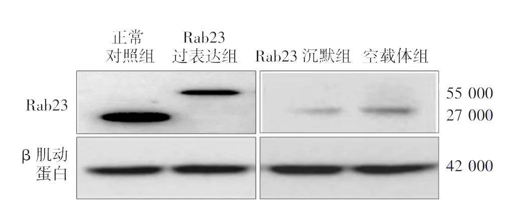
圖1 Western印跡檢測Sa3細胞Rab23表達水平 過表達Rab23組融合蛋白轉染成功,Rab23沉默組較空載體組相比,Rab23表達水平降低
表1 Rab23對Sa3細胞周期的影響(±s)
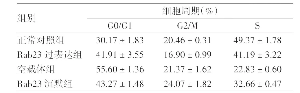
表1 Rab23對Sa3細胞周期的影響(±s)
注:n=3
組別 細胞周期(%)G0/G1 G2/M S正常對照組 30.17±1.83 20.46±0.31 49.37±1.78 Rab23過表達組 41.91±3.55 16.90±0.99 41.19±3.22空載體組 55.60±1.36 21.37±1.62 22.83±0.60 Rab23沉默組 43.27±1.48 24.07±1.82 32.66±0.47
2.平板克隆形成實驗證實Rab23抑制鱗癌Sa3細胞增殖:Rab23過表達組克隆形成率(2.3%±0.2%)較正常對照組(3.6%±0.3%)降低(t=4.02,P<0.05),Rab23沉默組(4.1%±0.2%)較空載體組(1.8%±0.0%)明顯增高(t=10.00,P<0.01)。提示Rab23對Sa3細胞增殖具有抑制作用。見圖2。
3.流式細胞儀檢測顯示Rab23抑制鱗癌Sa3細胞的增殖:見表1。Rab23過表達組細胞增殖指數(0.581±0.035)較正常對照組(0.698±0.018)降低(t=2.94,P<0.05),穩定轉染Rab23沉默組(0.567±0.015)較空載體組(0.444±0.014)顯著增高(t=6.12,P<0.01)。表明過表達Rab23可引起Sa3細胞G1期阻滯,影響細胞周期進程。
4.Sa3細胞Erk/p-Erk表達水平:Rab23過表達和Rab23沉默的Sa3細胞Erk蛋白水平較正常對照組和空載體組均無明顯變化。與正常對照組相比,Rab23過表達的Sa3細胞p-Erk蛋白水平降低,而Rab23沉默的Sa3細胞p-Erk蛋白水平較空載體組明顯增高。見圖3。
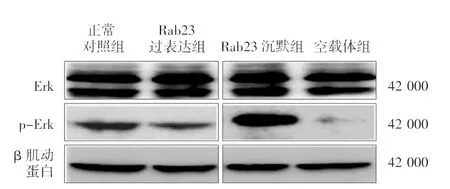
圖3 Western印跡檢測Erk/p-Erk表達水平

圖2 平板克隆實驗檢測Rab23對Sa3細胞增殖的影響 Rab23過表達組克隆形成率較正常對照組降低,Rab23沉默組較空載體組增高
討 論
Eggenschwiler等[9]發現,Rab23是Sonic Hedgehog(Hh)信號通路重要的負調控因子,Rab23作用位點在Hh信號通路的SMO和Gli分子之間,通過組織SMO分子進入纖毛而負調控Hh信號通路[10]。研究發現,在甲狀腺癌惡性類型中Rab23的mRNA水平低于濾泡狀腺瘤[11];在乳腺癌細胞中,過表達Rab23可降低Hh信號通路的激活狀態,并且抑制乳腺癌細胞的增殖[7]。而隨著研究的廣泛,發現Rab23在不同腫瘤細胞中對生長進程的影響并不一致,甚至呈相反的作用。在胃癌組織中,與非轉移型胃癌細胞相比,Rab23顯著高表達于轉移型胃癌細胞,研究者通過比較基因組雜交和基因表達分析將Rab23確定為胃癌侵襲介導因素;在肝癌組織中,Rab23表現為核陽性,其陽性率與腫瘤尺寸大小成正比,RNA干涉Rab23表達后癌細胞增殖能力下降,說明Rab23可促進肝癌細胞的增殖[6]。
侵襲性研究表明,Rab23促進鱗癌細胞的侵襲[8]。與此同時,Rab23對鱗癌細胞生長增殖的作用尚不清楚,因此我們首先通過慢病毒感染,構建穩定過表達、敲除Rab23的鱗癌Sa3細胞株。細胞周期檢測發現,Rab23可以引起鱗癌Sa3細胞G1期阻滯而阻礙細胞周期進程;平板克隆形成實驗發現,過表達Rab23組與對照組相比,Sa3細胞增殖水平降低,而敲除Rab23的Sa3細胞增殖能力提高,表明Rab23抑制鱗癌Sa3細胞增殖,這與Rab23對乳腺癌細胞生長的作用一致。
目前研究已明確,Hh信號通路過度激活可引發多種腫瘤。研究者通過免疫組化和原位雜交的方法,已明確在口腔和咽鱗癌[12]、肺鱗癌[5]組織、細胞中發現Hh信號通路的激活,并參與腫瘤的侵襲和生長。MAPK/Erk信號級聯反應也是影響多種癌細胞生長增殖的重要信號通路,可被表皮生長因子受體(EGFR)、整合素(integrins)、離子通道等激活。在多種上皮源性惡性腫瘤如頭頸部鱗癌細胞中,均有EGFR表達異常,并在鱗癌細胞的生長、侵襲、血管形成中起關鍵作用[13]。Morozevich等[14]發現,在皮膚鱗癌細胞中,EGFR-Erk通路的激活可促進鱗癌細胞增殖。而最近研究發現,在人髓母細胞瘤細胞中,Hh信號通路和EGFR信號通路存在交互作用[15]。我們初步研究發現,過表達Rab23的Sa3細胞,p-Erk表達水平較對照組降低;敲除Rab23的Sa3細胞,p-Erk表達水平較對照組升高,提示Rab23抑制鱗癌Sa3細胞增殖的機制可能與MAPK/Erk通路相關。而作為Hh信號通路必要的負調控因子,Rab23對鱗癌細胞增殖的抑制作用是單純通過MAPK/Erk通路還是同時通過Hh與EGFR信號通路有待進一步研究。
[1]Schaaij-Visser TB,Brakenhoff RH,Leemans CR,et al.Protein biomarker discovery for head and neck cancer[J].J Proteomics,2010,73(10):1790-1803.
[2]Barakat MT,Humke EW,Scott MP.Learning from Jekyll to control Hyde:Hedgehog signaling in development and cancer[J].Trends Mol Med,2010,16(8):337-348.
[3]Hou Q,Wu YH,Grabsch H,et al.Integrative genomics identifies Rab23 as an invasion mediator gene in diffuse-type gastric cancer[J].Cancer Res,2008,68(12):4623-4630.
[4]Sun HJ,Liu YJ,Li N,et al.Sublocalization of Rab23,a mediator of Sonic hedgehog signaling pathway,inhepatocellular carcinoma cell lines[J].Mol Med Rep,2012,6(6):1276-1280.
[5]Huang S,Yang L,An Y,et al.Expression of hedgehog signaling molecules in lung cancer[J].Acta Histochem,2011,113(5):564-569.
[6]Liu YJ,Wang Q,Li W,et al.Rab23 is a potential biological target for treating hepatocellular carcinoma[J].World J Gastroenterol,2007,13(7):1010-1017.
[7] 曾超,劉亞莉,胡玉珍,等.Rab23對乳腺癌細胞生長增殖的抑制作用[J].現代生物醫學進展,2009,9(20):3811-3815.
[8] 唐利,劉博強,簡強,等.Rab23在鱗癌細胞系中的表達及其對鱗癌細胞侵襲的影響[J].現代生物醫學進展,2012,12(13):2440-2443.
[9]Eggenschwiler JT,Espinoza E,Anderson KV.Rab23 is an essential negativeregulatorofthe mouse Sonic hedgehog signaling pathway[J].Nature,2001,412(6843):194-198.
[10]Boehlke C,Bashkurov M,Buescher A,et al.Differential role of Rab proteins in ciliary trafficking:Rab23 regulates smoothened levels[J].J Cell Sci,2010,123(Pt 9):1460-1467.
[11]Denning KM,Smyth PC,Cahill SF,et al.A molecular expression signature distinguishing follicular lesions in thyroid carcinoma using preamplification RT-PCR in archival samples[J].Mod Pathol,2007,20(10):1095-1102.
[12]Leovic D,Sabol M,Ozretic P,et al.Hh-Gli signaling pathway activity in oral and oropharyngeal squamous cell carcinoma[J].Head Neck,2012,34(1):104-112.
[13]Kalyankrishna S,Grandis JR.Epidermal growth factor receptor biology in head and neck cancer[J].J Clin Oncol,2006,24(17):2666-2672.
[14]Morozevich GE,Kozlova NI,Ushakova NA,et al.Integrinα5β1 simultaneously controls EGFR-dependent proliferation and Aktdependent pro-survival signaling in epidermoid carcinoma cells[J].Aging(Albany NY),2012,4(5):368-374.
[15]G?tschel F,Berg D,Gruber W,et al.Synergism between Hedgehog-GLIand EGFR signaling in Hedgehog-responsive human medulloblastoma cells inducesdownregulation ofcanonical Hedgehog-target genes and stabilized expression of GLI1[J/OL].PLoS One,2013,8(6):e65403[2013-09-30].http://www.plosone.org/article/info%3Adoi%2F10.1371%2Fjournal.pone.0065403.
2014-01-16)
(本文編輯:尚淑賢)
Inhibitory effect of Rab23 on the proliferation of a squamous cell carcinoma cell line Sa3 and its mechanisms
Liu Xilin,Jian Qiang,Miao Ye,Huang Min,Li Chengxin.Department of Dermatology,Xijing Hospital,Fourth Military Medical University,Xi'an 710032,China
Li Chengxin,Email:chengxinderm@163.com
ObjectiveTo evaluate the effect of Rab23 on the proliferation of a squamous cell carcinoma cell line Sa3,and to investigate its mechanisms.MethodsCultured Sa3 cells were classified into four groups:normal control group transfected with green fluorescent protein(GFP),Rab23-overexpressing group transfected with a GFP-labelled Rab23-overexpressing plasmid,Rab23-silencing group transfected with a plasmid carrying a Rab23-targeting shRNA,empty vector group transfected with an empty vector.After additional culture for different durations,plate colony formation assay and flow cytometry were performed to evaluate the proliferative activity of Sa3 cells,and Western blot was conducted to detect the expression of Erk/phosphorylated-Erk in Sa3 cells.Statistical analysis was carried out by t test,one-way analysis of variance and Bonferroni's multiple comparison test.ResultsStable Sa3 cell lines with overexpression or silencing of Rab23 were established by plasmid construction andlentivirus-mediated transfection.The plate colony formation assay showed that the colony formation rate was significantly lower in the Rab23-overexpressing group than in the normal control group(2.3%±0.2%vs.3.6%±0.3%,P<0.05),but higher in the Rab23-silencing group than in the empty vector group(4.1%±0.2%vs.1.8%±0.03%,P<0.01).Rab23 overexpression induced G1 phase arrest in Sa3 cells.The proliferation index was significantly decreased in the Rab23-overexpressing group compared with the normal control group(0.581±0.035 vs.0.698±0.018,P<0.05),but increased in the Rab23-silencing group compared with the empty vector group(0.567±0.015 vs.0.444±0.014,P<0.01).As Western blot showed,there were no significant changes in the expression of Erk in the Rab23-silencing or-overexpressing group compared with the normal control group,whereas the expression of p-Erk was attenuated in the Rab23-overexpressing group compared with the normal control group,but enhanced in the Rab23-silencing group compared with the empty vector group.ConclusionsRab23 could inhibit the proliferation of Sa3 cells,which may be associated with the Erk pathway.
Rab23;Lentivirus infections;Carcinoma,squamous cell;Cell line,tumor;Cell proliferation
10.3760/cma.j.issn.0412-4030.2014.07.014
710032西安,第四軍醫大學西京皮膚醫院
李承新,Email:chengxinderm@163.com
猜你喜歡
中國設備工程(2022年12期)2022-07-11 04:33:00
中學生數理化·七年級數學人教版(2021年6期)2021-11-22 07:50:58
中學生數理化·七年級數學人教版(2021年6期)2021-11-22 07:50:58
中學生數理化·七年級數學人教版(2021年6期)2021-11-22 07:50:58
中學生數理化·七年級數學人教版(2020年12期)2021-01-18 06:57:46
中學生數理化·七年級數學人教版(2020年12期)2021-01-18 06:57:46
中學生數理化·七年級數學人教版(2019年9期)2019-11-25 07:34:36
中學生數理化·七年級數學人教版(2019年9期)2019-11-25 07:34:34
中學生數理化·七年級數學人教版(2019年12期)2019-05-21 02:53:50
中學生數理化·七年級數學人教版(2019年12期)2019-05-21 02:53:48