Protective effect of camellia oil on H2O2-induced oxidative stress injury in H9C2 cardiomyocytes of rats
2024-03-26 12:55:08YANQingGUOZhenSUNSainanLIJingTANJiyongLIJing
YAN Qing, GUO Zhen, SUN Sai-nan, LI Jing, TAN Ji-yong, LI Jing
Department of Physiology, Guangxi Medical University, Nanning 530021, China
Keywords:
ABSTRACT Objective: To explore the protective effect of camellia oil against H2O2-induced oxidative stress injury in rat H9C2 cardiomyocytes.Methods: CCK8 method was used to detect the cell survival rate of H9C2 cardiomyocytes treated with different concentrations of H2O2.Normal cultured cells were used as the blank control group, and the cells were treated with 200 μmol/L H2O2 for 24 h.An oxidative stress injury model was constructed as the model group.The cells were pretreated with 1%, 0.1% and 0.01% camellia oil for 24 h, and then H2O2 was added for 24 h as the experimental group.The β-galactosidase senescence staining assay, mitochondrial membrane potential assay, EdU cell proliferation staining assay and scratch assay were used to observe the changes of cell senescence, mitochondrial membrane potential, proliferation,apoptosis and migration in each group.The superoxide dismutase (SOD) activity, lactate dehydrogenase (LDH) activity, and malondialdehyde (MDA) content of the cells in each group were detected by using the kit.Results: The cell viability of H9C2 cardiomyocytes treated with different concentrations of H2O2 was inhibited and positively correlated with the concentration of H2O2 (P<0.01).Compared with the blank control group, the positive rate of cell senescence,MDA content and LDH activity increased in the H2O2 model group (P<0.01); mitochondrial membrane potential, cellular value-added rate, migration rate and SOD activity decreased(P<0.01).Compared with the H2O2 model group, the positive rate of cellular senescence (P<0.01 or P<0.05), MDA content and LDH activity decreased (P< 0.01 or P<0.05); mitochondrial membrane potential increased, cell proliferation rate and migration rate increased (P<0.01 or P<0.05) in the experimental group.Conclusion: Camellia oil can significantly inhibit oxidative stress injury in H9C2 cells and exert cardiomyocyte protective effects.
1.Introduction
Globally Cardio Vascular Disease (CVD) is a disease with a long latency period, insidious onset, and difficult recovery that threatens human health[1].With the gradual aging of our society,CVD occupies the first place of mortality and has become one of the serious problems to be faced by today’s society[2].Oxidative stress refers to the disruption of intracellular reduction-oxidation imbalance through damage to lipids, DNA, and proteins when intracellular reactive oxygen species (ROS) are overproduced or antioxidants are depleted.Several cardiovascular diseases are associated with oxidative stress, such as atherosclerosis, congestive heart failure, ischemia-reperfusion injury, chronic ischemic heart disease, and arrhythmias[3].Hydrogen peroxide (H2O2) is a member of reactive oxygen species and as a messenger molecule, H2O2has a regulatory role in metabolism and it is also an important molecule in mediating oxidative stress in cells[4].Modelling oxidative stress using H2O2 induced cells has been widely used in experiments.
Camellia oil is widely used as an edible oil in China and is also considered as a complementary medicine.Camellia oil has a wide range of biological activities such as anti-inflammatory, anti-tumour,anti-hypertensive, antibacterial and protective effects against oxidative stress[5].However, there are few reports on the effects of camellia oil on cardiomyocytes.In this study, we used H9C2 cardiomyocyte cell line to establish an in vitro model to simulate H2O2-induced oxidative stress injury, and observed the protective effects of different concentrations of camellia oil on H2O2-induced oxidative injury in H9C2 cardiomyocytes.
2.Materials and methods
2.1 Materials
H9C2 cardiomyocytes (Pnocel LTD.); 3% H2O2(Sigma Corporation); Camellia oil (Shaanxi Jinkangtai Biotechnology Co.,LTD.); Dimethylsulfoxide (DMSO) (Sigma Company); CCK8 kit(New Saimei Biotechnology Co., LTD.) Fetal bovine serum (Gibco,USA); DMEM High sugar medium (Biological Industries, Israel);Pancrease (Israel Biological Industries); PBS (Soleibao Technology Limited); Streptomycin (Israel Biological Industries); Cell senescence β-galactosidase staining kit (Biyuntian Biotechnology Co., LTD.); BeyoClick? EdU-594 Cell Proliferation detection kit(Biyuntian Biotechnology Co., LTD.); Mitochondrial membrane potential detection Kit (JC-1) (Biyuntian Biotechnology Co., LTD.);LDH test kit, MDA test kit (Solaibao Co., LTD.); SOD detection kit(Nanjing Jiancheng Co., LTD.).
2.2 Methods
2.2.1 Cell culture
Rat H9C2 cardiomyocytes were cultured using DMEM highglucose medium containing 10% fetal bovine serum and 1%penicillin, and placed in a constant temperature incubator at 5%CO2and 37 ℃.The fluid was changed regularly, and when the cell density reached 80%, trypsin digestion was used for passaging.Cells in logarithmic growth phase were taken for experiments.
2.2.2 Preparation of camellia oil at different concentrations
Dissolve 10μl of camellia oil in 90l of DMSO to get 10%concentration of camellia oil; take 10l of 10% concentration of camellia oil and dissolve it in 90 μL of DMSO to get 1%concentration of camellia oil; dissolve it in this way in a gradient in order to get 10%, 1%, 0.1%, 0.01% concentration of camellia oil,respectively, in this study, we selected the three concentrations of 0.01%, 0.1%, and 1% to conduct the experiment.camellia oil for the experiment.
2.2.3 Cell grouping
H9C2 cells were inoculated in six-well plates at a density of 40,000 cells per ml and cultured for 24 h.After 24 h of culture, the cells were pretreated with camellia oil at concentrations of 1%, 0.1% and 0.01% for 24 h, and then treated with 200 μmol/LH2O2solution for 24 h.Cells were divided into the following groups: the blank control group, the model group: the H2O2group, and the experimental group: the H2O2+0.01% camellia oil group, H2O2+0.1% camellia oil group, H2O2+1% camellia oil group.Each group was set up with three compound holes, and the experiment was repeated three times.
2.3 Detection index
2.3.1 Detection of cell viability by CCK-8 method
H9C2 cells in the logarithmic growth phase were inoculated in ninety-six-well plates at a density of 40,000 cells per well, and after 24 h of normal cell culture, H2O2solution at concentrations of 0, 50,100, 200 and 400 μmol/L was added, and complete medium was added to the normal group.After 24 h of further incubation, 10 μL of CCK-8 solution was added, and the cells were incubated for 2 h at 37 ℃, the optical density (OD) value of each well was detected at 450 nm wavelength, and the cell survival rate was calculated.
2.3.2 β-galactosidase senescence staining experiment
After the cell culture solution was aspirated and washed once with PBS, add 1 mL of β-galactosidase staining fixative and fix it at room temperature for 15 minutes.The cells were washed with PBS after aspirating the fixative.Add 1mL of β-galactosidase staining working solution to each well and incubate in a constant temperature incubator at 37 ℃ without CO2overnight.Cell staining was observed under an ordinary light microscope, senescent cells were green and non-senescent cells were not coloured.The results were expressed as the percentage of senescent staining positive cells in the total counted cells.
2.3.3 Mitochondrial membrane potential assay
Aspirate the cell culture medium, wash once with PBS, add 1 mL of culture medium and 1 mL of JC-1 staining working solution to each well, mix thoroughly.Incubate at 37 ℃ for 20 min.At the end of incubation, wash twice with JC-1 staining buffer (1×).Add 2ml cell culture solution and observe the cells under fluorescence inverted microscope.Mitochondrial membrane potential fluorescence intensity was analysed using Image J software.The ratio of red/green fluorescence intensity was used to indicate the degree of cell depolarisation, and the high or low ratio represented the high or low membrane potential.
2.3.4 EdU cell proliferation staining experiment
Add EdU working solution into cells and incubate for 2 h to complete EdU labelling.The cells were incubated with Click Additive Solution at room temperature and protected from light for 30 min, and washed at the end of the incubation, and incubated with 1× Hoechst 33342 solution at room temperature and protected from light for 10 min.After washing, the cells were observed under a fluorescence inverted microscope.The cell proliferation rate was calculated by ImageJ software.
2.3.5 Scratch experiment
Discard the cell culture solution, use a ruler and 200 μL gun tip to make scratches perpendicular to the six-well plate, make three equally spaced vertical lines in each well, add PBS to wash the cells to remove the cell debris after scratches are made, and add the culture medium.Photographs were taken with an ordinary light microscope to record the migration of cells at the time of first scratching and after 24 h.The cell migration rate was also calculated by ImageJ software.
2.3.6 Detection of LDH, MDA and SOD levels
Collect the cells and supernatants of each group, and test the levels of LDH and SOD in the supernatants, and the levels of MDA in the cells according to the instructions of the LDH, MDA and SOD assay kits, respectively.
2.4 Statistical processing
SPSS 23.0 and GraphpadPrism 8.0 statistical software were used for data analysis.The experimental data were expressed as means± standard deviation , and t-test or one-way ANOVA was used for intergroup comparisons, and P < 0.05 was considered to statistically significant difference.
3.Results
3.1 Establishment of H2O2 oxidative stress injury model
The H2O2-induced H9C2 cell injury model was established to simulate the damage to cardiomyocytes during oxidative stress.The results showed that compared with the blank control group,the viability of H9C2 cells decreased with the increase of H2O2concentration, and the viability of H9C2 cells decreased to about 52% of that of the control group after treating the cells with 200 μmol/L H2O2for 24 h (P<0.01), which indicated that this concentration could significantly inhibit the viability of H9C2 cells without causing a large number of apoptosis, so the subsequent experiments were carried out using 200 μmol/L H2O2treatment of cells for 24 h to establish the cell injury model, Table 1.
Tab 1 Comparison of cellular activity after H9c2 cardiomyocytes intervention with different concentrations of H2O2(n=5, ±s)

Tab 1 Comparison of cellular activity after H9c2 cardiomyocytes intervention with different concentrations of H2O2(n=5, ±s)
Note:vs.0mol/L group, *P<0.05; **P<0.01.
H2O2(μmol/L) survival rate(%)0 100 50 83.31±0.47**100 75.69±0.30**200 52.80±0.31**400 12.58±0.50**F 34178 P 0.000
3.2 Effects of camellia oil on senescence of H9C2 cells after H2O2 injury
The results showed that compared with the blank control group,the positive rate of β-galactosidase in the model group increased,and the percentage of senescent cell number increased (P<0.01);compared with the model group, the number of senescent cells in the experimental group decreased, the percentage of senescent cell number decreased, and with the increase of the concentration of camellia oil, the number of senescent cells sequentially decreased,and the positive rate of senescent cells decreased (P<0.01 or P<0.05), Table 2, Figure 1.
Tab 2 Effect of camelliaoil on the positive rate of H2O2-induced senescence in H9C2 cells(n=3, ±s)
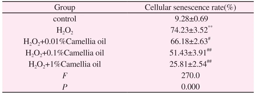
Tab 2 Effect of camelliaoil on the positive rate of H2O2-induced senescence in H9C2 cells(n=3, ±s)
Note:vs.control group, **P<0.01; vs.H2O2 group, #P<0.05; ##P<0.01.
Group Cellular senescence rate(%)control 9.28±0.69 H2O2 74.23±3.52**H2O2+0.01%Camellia oil 66.18±2.63#H2O2+0.1%Camellia oil 51.43±3.91##H2O2+1%Camellia oil 25.81±2.54##F 270.0 P 0.000
3.3 Effects of camellia oil on mitochondrial membrane potential of H9C2 cells after H2O2 injury
The results showed that compared with the blank control group, the ratio of cellular red/green fluorescence intensity in the model group was significantly reduced (P<0.01); compared with the model group,the ratio of cellular red/green fluorescence intensity in theH2O2+1%camellia oil group was elevated (P<0.01); there was no significant difference in the cellular red/green fluorescence intensity between the H2O2+0.1% camellia oil group, H2O2+0.01% camellia oil group(P>0.05), Figure 2.
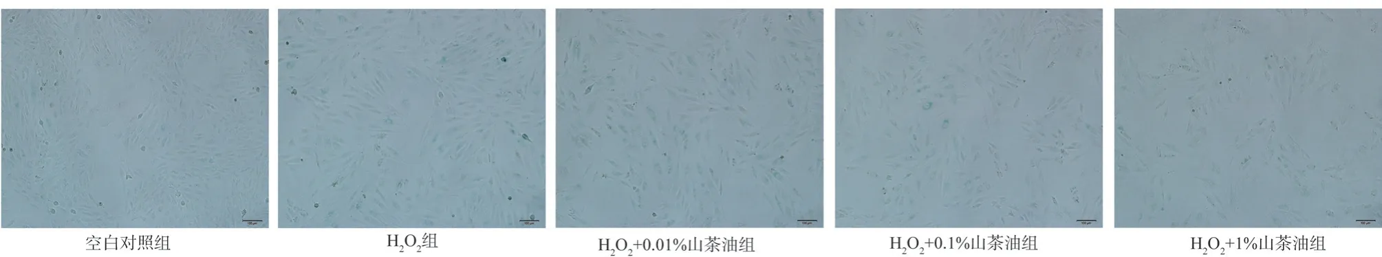
Fig 1 Senescence-associated-β-galactosidase (SA-β-gal) staining of H9C2 cells after different concentrations of camellia oil intervention model(×100)
3.4 Effect of camellia oil on the proliferation of H9C2 cells after H2O2 injury
The results showed that compared with the blank control group, the cell proliferation rate of the model group was significantly reduced(P<0.01); compared with the model group, the cell proliferation rate of the experimental group increased, and the proliferation rate increased sequentially with the increase of the concentration of camellia oil (P<0.01 or P<0.05), Table 3, Figure 3.

Fig 2 Mitochondrial membrane potential assay (JC-1) of H9C2 cells after different concentrations of camellia oil intervention model(×100)
Tab 3 Effectofcamellia oil on the proliferation rate of H2O2-induced H9C2 cells(n=3,±s)
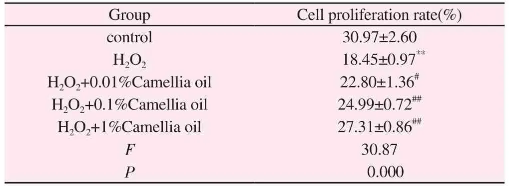
Tab 3 Effectofcamellia oil on the proliferation rate of H2O2-induced H9C2 cells(n=3,±s)
Note:vs.control group, **P<0.01; vs.H2O2 group, #P<0.05; ##P<0.01.
Group Cell proliferation rate(%)control 30.97±2.60 H2O2 18.45±0.97**H2O2+0.01%Camellia oil 22.80±1.36#H2O2+0.1%Camellia oil 24.99±0.72##H2O2+1%Camellia oil 27.31±0.86##F 30.87 P 0.000
3.5 Effect of camellia oil on the migration of H9C2 cells after H2O2 injury
The results showed that compared with the blank control group,the cell migration rate of the model group decreased and the healing ability was weakened (P<0.01); compared with the model group,there was no significant difference in the migration rate between the H2O2+0.01% camellia oil groups, and the cell migration rate increased and the cell healing ability was enhanced in the H2O2+0.1% camellia oil group and the H2O2+1% camellia oil group(P<0.01 or P<0.05).Table4, Figure 4.
Tab 4 Effect of camellia oil on H2O2-induced migration rate of H9C2 cells(n=3,±s)
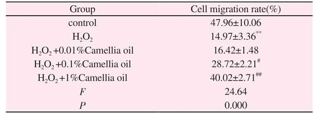
Tab 4 Effect of camellia oil on H2O2-induced migration rate of H9C2 cells(n=3,±s)
Note: vs.control group, **P<0.01; vs.H2O2 group, #P<0.05; ##P<0.01.
Group Cell migration rate(%)control 47.96±10.06 H2O2 14.97±3.36**H2O2 +0.01%Camellia oil 16.42±1.48 H2O2 +0.1%Camellia oil 28.72±2.21#H2O2 +1%Camellia oil 40.02±2.71##F 24.64 P 0.000
3.6 Effects of camellia oil on LDH, MDA and SOD in H9C2 after H2O2 injury
Compared with the blank control group, cellular SOD activity in the model group decreased (P<0.01), LDH activity and MDA content increased (P<0.01); compared with the model group, there was no significant difference in MDA content and SOD activity and an increase in LDH activity in the cardiomyocytes between the H2O2+0.01% camellia oil groups (P<0.01).Cellular SOD activity increased and LDH activity decreased in the H2O2+0.1% camellia oil group and H2O2+1% camellia oil group (P<0.01).Cellular SOD activity increased, LDH activity and MDA content decreased in the H2O2+1% camellia oil group.1% camellia oil group showed an increase in cellular SOD activity and a decrease in LDH activity and MDA content (P<0.01 or P<0.05).Table 5.

Fig 3 EdU staining of H9C2 cells after different concentrations of camellia oil intervention model(×200)
Tab 5 Effect of camellia oil on LDH, MDA and SOD in H2O2-induced H9C2 cells(n=3, ±s)
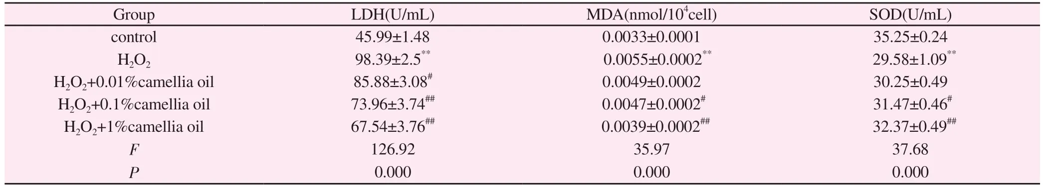
Tab 5 Effect of camellia oil on LDH, MDA and SOD in H2O2-induced H9C2 cells(n=3, ±s)
Note: vs.control group, **P<0.01; vs.H2O2 group, #P<0.05;##P<0.01.
Group LDH(U/mL) MDA(nmol/104cell) SOD(U/mL)control 45.99±1.48 0.0033±0.0001 35.25±0.24 H2O2 98.39±2.5** 0.0055±0.0002** 29.58±1.09**H2O2+0.01%camellia oil 85.88±3.08# 0.0049±0.0002 30.25±0.49 H2O2+0.1%camellia oil 73.96±3.74## 0.0047±0.0002# 31.47±0.46#H2O2+1%camellia oil 67.54±3.76## 0.0039±0.0002## 32.37±0.49##F 126.92 35.97 37.68 P 0.000 0.000 0.000
4.Discussion
Cardiovascular diseases include arrhythmias, coronary atherosclerotic heart disease, hypertension, cardiomyopathy,peripheral vascular disease and congenital heart defects[6,7].Cardiovascular disease is the main cause of death in China,accounting for 40% of the death of China’s population, is deservedly the “first killer” of human health[8].With the gradual increase in the proportion of the elderly population in China, the incidence of cardiovascular disease is also increasing, we must pay attention to its prevention and treatment.
Many chronic diseases, including cardiovascular diseases, such as diabetes, renal and skeletal muscle diseases, have been shown to be directly related to oxidative stress[9].Oxidative stress refers to an imbalance between oxidative and antioxidant mechanisms in the body, and reactive oxygen species is a collective term for a class of chemically reactive oxygen-containing chemicals,including perhydroxyl radicals superoxide, oxides, and other nonradical molecules.Reactive oxygen species levels are critical for the regulation of cellular homeostasis, and low to moderate levels of reactive oxygen species activate stress-responsive survival pathways that maintain cell differentiation and proliferation.However,high levels of reactive oxygen species cause damage to cellular components such as DNA, proteins and lipids[10].A variety of cardiovascular diseases have been shown to be associated with the overproduction of reactive oxygen species[11], H2O2 as a member of reactive oxygen species regulates a variety of biological processes ranging from cell differentiation to apoptosis, while excess H2O2 is also widely recognised as having cytotoxic effects, which are eliminated in vivo by enzymes such as catalase and glutathione peroxidase[12,13].
Camellia oil is a purely natural edible oil obtained from mature Camellia sinensis seeds and is often referred to as “Asian olive oil”because of its high concentration of oleic acid, which is very similar to olive oil.Camellia oil is not only a common edible oil, but also has great medicinal value.Traditionally, camellia oil has long been used to treat people with gastrointestinal, lung and kidney diseases.Previous studies have shown that camellia oil is more effective in preventing hypertension, hyperlipidaemia and hyperglycaemia than common cooking oil[14,17].The main components of camellia oil include oleic acid, linoleic acid, palmitic acid, squalene, vitamin E,and flavonoids, which have a wide range of pharmacological effects,including antioxidant effects.From this, we conjecture that camellia oil can alleviate cardiovascular damage caused by oxidative stress,thus effectively preventing cardiovascular disease.
The use of H2O2 to induce cellular oxidative stress injury model in vitro has gradually matured, and this study explores the protective effect of camellia oil on cellular oxidative stress by using H2O2to establish a cellular oxidative stress injury model in vitro with an oxidative stress injury model.Firstly, H9C2 cardiomyocytes were modelled with different concentrations of H2O2, and the optimal H2O2damage modelling concentration was selected by CCK8 experiment to establish the oxidative stress model.After pre-treating the cells with different concentrations of camellia oil,we added 200 mol/L H2O2to stimulate the cells, and explored the effects of different concentrations of camellia oil on cell senescence, mitochondrial membrane potential, cell proliferation,cell migration and oxidative stress indicators after oxidative stress through the β-galactose staining, mitochondrial membrane potential, cell proliferation, and the scratch assay respectively.indicators of oxidative stress.The results of this study showed that under the condition of H2O2treatment, camellia oil could inhibit the decrease of mitochondrial membrane potential and senescence of cardiomyocytes, and increase the proliferation and migration of cardiomyocytes.LDH and MDA could reflect the degree of oxidative damage of cardiomyocytes, and SOD could reflect the degree of antioxidant of cardiomyocytes.Camellia oil can reduce the LDH and MDA activities of cardiomyocytes under the condition of H2O2treatment and increase the SOD activity.This indicates that camellia oil can play a protective role against H2O2-induced H9C2 cardiomyocyte injury.
In conclusion, the present study confirmed the protective effect of camellia oil against H2O2-induced oxidative stress injury in rat H9C2 cardiomyocytes, suggesting that camellia oil may have the ability to resist oxidative stress injury and that the antioxidant power of camellia oil is enhanced with the increase of camellia oil concentration.The experimental results provide help for the subsequent antioxidant pharmacological effects of camellia oil and clinical studies.The present study was limited to in vitro cellular experiments only, and further validation in animal models is needed to provide more evidence for the protective effect of camellia oil against oxidative damage in the organism.
Authors’ contribution
Yan Qing: experimental implementation, data processing and paper writing; Nicco Guo, Sai-Nan Sun and Jing Lai: literature search and data analysis; Jing Lai and Kiyong Tan: experimental design,experimental guidance and paper revision and correction.
All authors declare no conflict of interest.
 Journal of Hainan Medical College2024年1期
- Journal of Hainan Medical College的其它文章
- Mechanism of traditional Chinese medicine in the treatment of psoriasis with depression: A review
- Molecular mechanisms of ferroptosis and its role in the treatment of breast cancer
- Research progress in mitochondrial autophagy mediated by BNIP3
- Meta analysis of the efficacy of western medicine combined with Qiliqiangxin capsule versus western medicine alone in the treatment of chronic heart failure
- Bioinformatics and network pharmacology identify the therapeutic role and potential targets of diosgenin in Alzheimer disease and COVID-19
- Pharmacodynamic study and mechanism of action of Linggui Zhugan Decoction in the intervention of Nonalcoholic fatty liver disease