Atypical Adams-Oliver syndrome with typical ocular signs of familial exudative vitreoretinopathy
2022-08-10 01:39:28EnZhongJinLyuZhenHuangMingWeiZhaoHongYin
INTRODUCTION
Adams-Oliver syndrome (AOS) is a rare inherited condition first described in 1945
. As a multiple malformation syndrome, AOS was characterized by a combination of aplasia cutis congenita (ACC) and variable degree of transverse limb defects
. Several genes associated with AOS including
and
have been reported in the past decade
. In previous studies, the systemic phenotype within families of AOS may range from no obvious clinical manifestations in mutation carriers to severe multiple-system anomalies that can even result in miscarriage or stillbirth
.
1.6 統計學處理 采用SPSS 17.0統計學軟件進行統計分析,以P<0.05為差異有統計學意義。計量資料以表示,計數資料以率表示。單因素分析采用Student′s t檢驗或四格表χ2檢驗,將P<0.1的因素納入多因素分析,多因素分析采用二項分類Logistic回歸模型,通過多因素分析控制混雜因素,明確胸腰椎結核手術患者早期植骨融合的獨立影響因素。
小規模納稅人的科目設置參照《規定》,在“應交稅費”科目下設置“應交增值稅”、“轉讓金融商品應交增值稅”、“代扣代交稅金”明細科目,核算原理同上。
MATERIALS AND METHODS
This study was approved by the Ethical Review Committee of Peking University People’s Hospital(Beijing, China), which was conducted in accordance with the Declaration of Helsinki. Written informed consent for genetic testing and medical photograph collection was obtained from the parents.
“兒童離不開生活,生活離不開健康教育。”晨間戶外鍛煉作為體育活動的活動形式之一不僅能讓幼兒有效地鍛煉身體,擁有健康,而且能更多直接地接受陽光、新鮮空氣和水分等自然因素的刺激,所以只有讓幼兒充分體驗晨間戶外體育鍛煉活動的快樂,形成活潑、向上的性格,才能更好地促進各領域的學習,才能提高幼兒的生活乃至生命的質量。
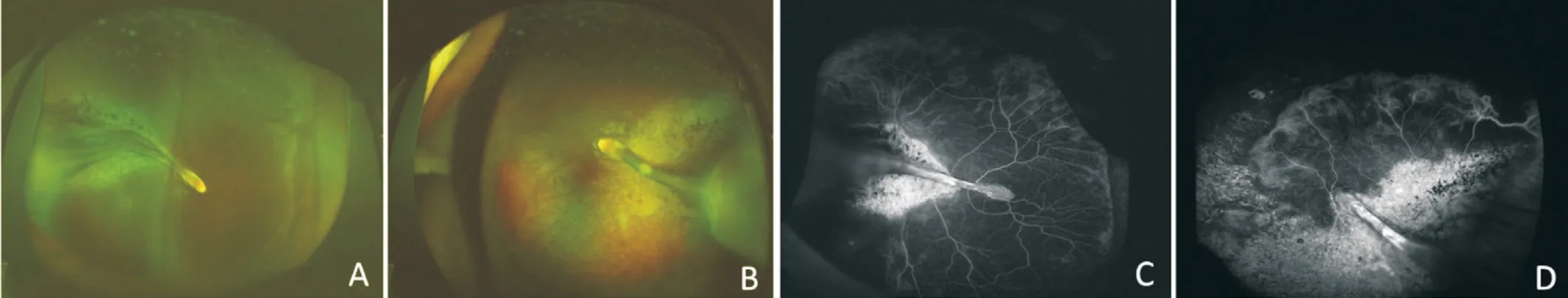
The boy born at full-term with birth weight of 3000 g, and no special prenatal history was recorded. The rough visual acuity of the boy was 20/80 for both eyes with poor cooperation. Noncontact intraocular pressure was 17 mm Hg and 16 mm Hg for right and left eye, respectively. The cycloplegic refraction was measured as follows: -0.25 D for the right eye and -4.00 D for the left eye. No obvious abnormalities can be observed for the examination of the anterior segment of the eyes. For the binocular indirect ophthalmoscope fundus examination, normal boundary and color of the optic discs can be showed, while the avascular area was visible in the periphery retina. Ultrawide-field FA examination was performed for the patient and obvious non-perfusion in the peripheral retina can be defined.Abnormal vessels can be observed at the junction of vascular and avascular retina though no leakage was identified (Figure 1).Fundus color photographs and FA images were also collected for the boy’s elder sister and parents. For the sister, significant peripheral retinal detachment can be observed in both eyes with temporal retinal folds originating from the optic disc, and the temporal retina was gray-white with hyperpigmentation sounded. FA examination showed peripheral retinal nonperfusion area and abnormal vascularization including tortuous veins, arteriovenous shunt, abnormal anastomosis. Retinal atrophy and hyperpigmentation can also be identified in fluorescein angiogram (Figure 2).
該方法根據網絡的不同情況將網絡進行拆分,再根據每一個部分的特點采用不同的方法進行混合預測.De[13]等人根據節點的屬性和網絡結構將目標網絡分為兩層,第一層根據節點獲得的信息判斷兩個節點之間產生連接的可能性,第二層根據網絡的結構,結合AA指標,采用協同聚類的方式獲取特征值,最后通過支持向量機完成整個預測過程;Chen[14]等人提出網絡稀疏化框架,通過四種不同的方法稀疏化目標網絡,對稀疏化后的網絡逐一訓練分類器,并整合在一起進行綜合預測.該方法將特定網絡以特定方法分解后能有效預測網絡鏈路,但普適性不佳.
Ophthalmologic examination was performed with a standard protocol including best-corrected visual acuity (BCVA), slit-lamp examination, binocular indirect ophthalmoscope fundus examination, ultra-wide-field fundus photography and fluorescein angiography (FA; Optos Daytona, Optos PLC, Dunfermline, UK). All the examination data was collected along with the data of medical history. All the digital images of fundus and FA were blinded reviewed by two experienced ophthalmologists (Yin H and Jin EZ).
Whole exome sequencing was performed to characterize mutations for the proband boy and all his family members. Peripheral blood samples were obtained for the gene sequencing and sent to an external service to be sequenced.
RESULTS
The genetic sequencing was performed for the whole family and the results indicated that the children (both the boy and his sister) had two compound heterozygous mutations (c.1396C>T p.R466X, c.4796G>A p.W1599X) in the gene
associated with AOS type 2, and the family verification results showed the two heterozygous mutations were originated from their father and mother, respectively. Pedigrees of the family and sanger confirmation of the identified
variants in proband with family members are shown in Figure 3.
The AOS associated with ocular findings is rarely reported especially for the patients initially diagnosed in Ophthalmology Department. Almost all reports associated with ocular disorders were cases. In a previous literature, a female AOS case with bilateral congenital cataract was reported
. In another report, a case of AOS mimicked as familial exudative vitreoretinopathy(FEVR) had been reported recently, but genetic testing was not performed in this single case
. In 2012, an AOS case presenting retinal findings consistent with ischemicproliferative retinopathy had also been reported
. Since AOS was found to be associated with several ocular disorders and few cases have already been reported, the relationship between AOS and ocular diseases worth further discussion.In our present report, a family of AOS with ocular signs of FEVR was identified according to the clinical and genetic findings with
mutation. The ophthalmic examinations and gene sequencing were performed for the patient, his elder sister and their parents.
A 5-year-old boy initially diagnosed with FEVR and found to have AOS according to the systemic situation, family history and gene sequencing was described. The individual and family investigation were performed. All family members of the proband boy including the parents and his 10-year-old sister were included in the present study and underwent ocular and genetic examinations for further analysis.
In this case, the parents of the boy showed no obvious ocular symptoms or signs, but the boy showed slightly lower intelligence, poor vision, mild esotropia and large nonperfusion zone at the temporal retina, which suggesting FEVR-like fundus changes. For the family history, it was found that the child’s sister had a history of retinal detachment with retinal fold in both eyes. Fundus examinations showed obvious retinal folds in both eyes, with small palpebral fissure,esotropia, raised outer corner of the eye, right hand penetrating palm, high muscle tension, mild hemiplegia of the left limb,
. There are many similarities between these manifestations and the previously reported systemic and ocular signs of AOS
.
The 5-year-old boy was found to have abnormal eye position and slightly mental retardation from birth according to his parents’ discription. No physical deformitie and mental abnormalities existed among his parents. For further family members’ investigation, his 10-year-old sister previously defined as retinal detachment and clinically diagnosed with FEVR. She was found to have poor vision, esotropia,blepharophimosis, upward of external eye corner, mild mental retardation and left hemiplegia.
DISCUSSION
AOS was not frequently reported in patients with ocular disorders though some isolated cases had been described associated with congenital cataract and retinal findings
. In our case, we described a family of AOS with all the members showing retinal signs similar to FEVR and
mutations.To the best of our knowledge, this is the first report of AOS family identified by gene mutations with mimicking FEVR retinal findings.
As we all know, AOS was first reported in 1945, mainly manifested as scalp defects and varying degrees of limb defects
.The AOS syndrome can be inherited in autosomal dominant manner in most cases, and in autosomal recessive manner for some cases (
and
gene mutations)
. The underlying pathological mechanism of AOS is considered as a congenital vascular disease that may involve multiple systems such as the cardiovascular system, brain, liver, lung,eyes and skin
. In the past, few reports about AOS related to ocular disorders, especially for retinal manifestations
.The insufficient evidence between AOS and retinal disorders like FEVR made the present study more meaningful. But the limitations of our study can not be ignored. First, only one family of AOS mimicked FEVR was included which may reduce the persuasion, more families or cases should be collected and combined analyzed. Second, as a report of family case, no functional validation was performed even though two novel mutations were found. Third, the ocular signs of FEVR of the proband and his sister was typical while the AOS signs of them were atypical, but the genetic sequencing can provide most strong evidence.


For their parents, the FA examinations were also performed.Only avascular zone and abnormal anastomosis at the vascularavascular junction can be identified for them and both of them can reached a BCVA of 20/20. Meanwhile, no physical deformitie and mental abnormalities existed among the parents.
Basing on the ocular and systemic manifestations of the boy and his sister, a whole-exome sequencing analysis of the single-gene disease was performed on the whole family. On one hand, further clarification whether there are FEVR-related genetic changes were performed, and on the other hand, it is also analyzed whether there was genetic mutation associated with AOS. The sequencing revealed that the proband had two heterozygous mutations (c.1396C>T p.R466X, c.4796G>A p.W1599X) in
which is associated with the AOS type 2. Though the variant in
was reported to be associated with AOS type 2 before, these two novel missense mutations identified in this study had not been previously registered in the Ensembl database (http://www.ensembl.org/index.html) or the Human Gene Mutation Database (HGMD, http://www.hgmd.cf.ac.uk/ac/index.php)
. And the compound heterozygous mutations of them were found to be associated with the AOS proband. The further family verification results showed that the two heterozygous mutations came from their parents respectively and were compound heterozygous mutations. The sister also carries the two compound heterozygous mutations.As truncating mutations, the two novel missense mutations show high potential pathogenicity. Our findings expand the mutational spectrum of
and suggest that the two compound heterozygous mutations of
is associated with AOS.
For the previous reports, the retinal fold with or without retinal detachment had been reported in AOS patients, and the retinal folds involving the macular was also described in one case
. A seven-week-old full-term infant within normal birth weight clinically diagnosed with AOS with ocular signs similar to FEVR was reported
. While no remarkable change existed in anterior segment, a radial falciform retinal fold extending from the macula to temporal periphery can be defined in this case. Significant preretinal fibrous proliferation can be noted, but no neovascularization or exudation were observed, and no ophthalmic examination was performed for the parents and three male siblings of the infant
. In our case,the whole family underwent gene sequencing and ophthalmic examinations, and genetic mutations can be identified accurately. Retinal detachment accompanied by retinal fold can also be found in the sister, along with poor vision, mild mental retardation and left hemiplegia. The ocular and systemic disorders of the sister confirmed by the genetic sequencing can further assist the diagnosis of AOS for the whole family.In this case, FEVR-like fundus and fluorescein angiogram can be observed in all family members. Despite the presence of FEVR-like changes existed, they were generally quiet. No obvious peripheral leakage or retinal traction can be observed,and the boy was only asked for closely and regular follow-up without treatment.
As an inherited retinal disorder characterized by abnormal development of retinal vasculature, FEVR was thought to be associated with Wnt signaling pathway
. Several genes including
, and
had been found to be associated with FEVR in previous studies
. On the other hand, these genes were also identified in atypical retinopathy of prematurity (ROP) patients
. The Wnt signaling pathway may play a common pathological role in them, which is a major role for the development of retinal vascular. According to the whole-exome sequencing analysis, no special mutations of these genes can be found in our present family case, which did not support the relationship between AOS and Wnt signaling pathway. Since no association between FEVR and
had been identified before, and the
and
involved in the Notch signaling pathway were found to play a crucial role in developing blood vessel walls, the pathogenesis of AOS may be different with FEVR
. Although a case of AOS with similar changes mimicking FEVR diagnosed basing on clinical systemic characteristics has been reported,the present case is the first confirmed family case basing on genetic sequencing. Our report provides information on genetic mutations and clinical features to assist the ophthalmologists in recognizing AOS patients. Also, this family case can help us further understanding the ocular manifestations of AOS. It can be defined that the ocular phenotype of AOS may mimic that of FEVR. The patients diagnosed with AOS should be further evaluated for the retinal vasculopathy by fundus examinations including fluorescein angiography to make sure whether specific treatment was required. And the FEVR patients should also be carefully evaluated for the potential possibility of suffering from AOS.
ACKNOWLEDGEMENTS
Jin EZ: Project development, data management and analysis, manuscript writing and editing;Huang LZ: Data analysis, manuscript editing; Zhao MW:Manuscript editing; Yin H: Project development, manuscript writing and editing.
2.1.1 子宮出血的特點:月經周期發生紊亂,出血量時少時多,經期的長短不定,有時有大量出血。出血期無下腹痛或其他不適,出血量多或時間長者可伴發貧血癥狀,大量出血時可導致休克[2]。
登錄APP查看全文
猜你喜歡
天津教育(2023年2期)2023-03-14 07:34:52
中老年保健(2022年5期)2022-08-24 02:36:04
當代陜西(2021年12期)2021-08-05 07:45:46
中國生殖健康(2020年7期)2021-01-18 03:02:24
甘肅教育(2020年6期)2020-09-11 07:45:12
經濟技術協作信息(2018年8期)2019-01-14 03:06:28
現代營銷(創富信息版)(2018年9期)2018-09-03 09:49:38
消費導刊(2017年24期)2018-01-31 01:29:28
冰雪運動(2016年4期)2016-04-16 05:54:56
劍南文學(2015年1期)2015-02-28 01:15:15
 International Journal of Ophthalmology
2022年8期
International Journal of Ophthalmology
2022年8期
- International Journal of Ophthalmology的其它文章
- Advances in the research of plant-derived natural products against retinoblastoma
- Acute bilateral anterior uveitis in paediatric inflammatory multisystem syndrome temporally associated with COVlD-19
- Metamorphopsia as the first clinical sign of renal cell carcinoma
- Sutureless contact lens-type amniotic membrane for persistent epithelial defects after infectious keratitis
- A case of conjunctival intraepithelial neoplasia with spheroidal degeneration: a clinicopathological study
- Risk of anxiety and depression in patients with uveitis: a Meta-analysis