上皮樣膠質(zhì)母細胞瘤診斷與顯微治療
2022-07-15 01:25:44孔騰霄張棟韜袁善鵬高鵬焦賀男李雪元閆東明
中國現(xiàn)代醫(yī)生
2022年16期
關(guān)鍵詞:磁共振成像
孔騰霄 張棟韜 袁善鵬 高鵬 焦賀男 李雪元 閆東明
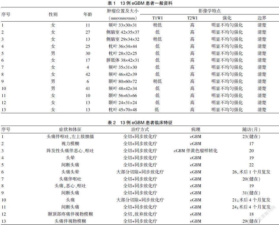
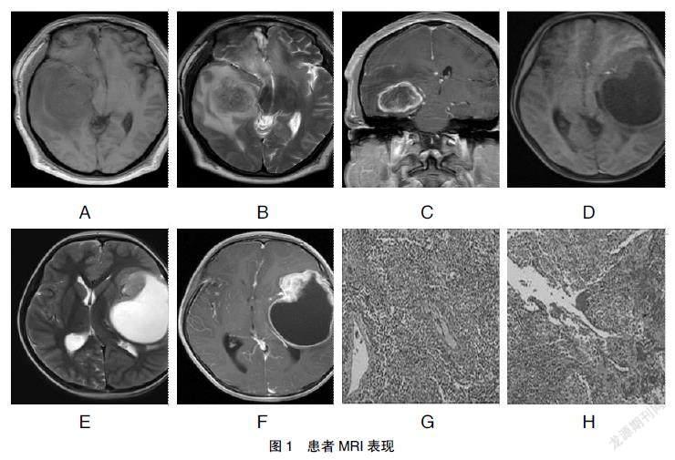
[摘要] 目的 探討上皮樣膠質(zhì)母細胞瘤(eGBM)的診斷方式、臨床特點、治療方法。 方法 回顧性分析2012年1月~2020年1月鄭州大學第一附屬醫(yī)院收治的13例eGBM患者的資料,其中男3例,女10例,年齡4~42歲,平均(19.4±12.5)歲。病變部位:額葉4例,枕葉3例,顳葉3例,側(cè)腦室2例,胼胝體1例,患者均行顯微手術(shù)治療。描述腫瘤臨床特征及影像、病理特點,鑒別分析與其相混淆病變,隨訪患者治療效果及預后情況。 結(jié)果 eGBM臨床表現(xiàn)多為因顱內(nèi)占位而引起的頭痛、嘔吐、視力模糊等癥狀;影像學檢查具有一定的特征性:瘤體實性部分T1WI為低/稍低信號,T2WI為高信號,增強掃描呈不均勻強化,發(fā)生部位表淺,囊變、壞死、出血多,邊界清楚,常累及鄰近腦膜且伴周圍組織水腫;11例術(shù)中全切,2例行大部分切除,12例術(shù)后同步放、化療。患者中位生存期為21個月(至末次隨訪仍有4例患者健在)。術(shù)后3例患者出現(xiàn)復發(fā)。免疫組化顯示CK、GFAP、S-100陽性率較高和Ki-67增殖指數(shù)的高表達;分子生物學方面常表達INI-1和B-raf基因V600E突變。結(jié)論 eGBM的臨床特點無特異性,一般為顱內(nèi)占位引起的顱高壓癥狀,其診斷依賴于放射學、組織學和遺傳學的綜合分析,顯微手術(shù)切除和同步放化療仍是一線治療方式。
[關(guān)鍵詞] 上皮樣膠質(zhì)母細胞瘤;磁共振成像;病理學;顯微治療
[中圖分類號] R739.4? ? ? ? ? [文獻標識碼] A? ? ? ? ? [文章編號] 1673-9701(2022)16-0010-05
Diagnosis and microsurgery of epithelioid glioblastoma
KONG Tengxiao? ?ZHANG Dongtao? ?YUAN Shanpeng? ?GAO Peng? ?JIAO He′nan? ?LI Xueyuan? ?YAN Dongming
Department of Neurosurgery, the First Affiliated Hospital of Zhengzhou University, Zhengzhou 450052, China
[Abstract] Objective To investigate the diagnosis method, clinical characteristics and treatment of epithelioid glioblastoma (eGBM). Methods The data of 13 patients with eGBM admitted to the First Affiliated Hospital of Zhengzhou University from January 2012 to January 2020 were retrospectively analyzed, including 3 males and 10 females, aged(19.4±12.5)years (range:4-42 years).The lesions were frontal lobe in 4 cases,occipital lobe in 3 cases,temporal lobe in 3 cases,lateral ventricle in 2 cases,and corpus callosum in 1 case.The clinical features,imaging and pathological features of the tumor were described. The confused lesions were differentiated and analyzed,and the therapeutic effect and prognosis of the patients were followed up. Results The clinical manifestations of eGBM were mainly headache, vomiting, blurred vision and other symptoms caused by intracranial mass.Imaging examination had certain characteristics:The solid part of the tumor was hypointense/slightly hypointense on T1WI,hyperintense on T2WI, heterogeneous enhancement on enhanced scan, the occurrence site was superficial,with cystic degeneration, necrosis, bleeding, clear boundary,often involving the adjacent meninges and with surrounding tissue edema.11 cases underwent total resection,2 cases underwent major resection, and 12 cases underwent postoperative concurrent radiotherapy and chemotherapy.The median survival time was 21 months(4 patients remained alive until the last follow-up).Postoperative recurrence occurred in 3 patients. Immunohistochemistry showed a high positive rate of CK,GFAP,and S-100 and a high expression of Ki-67 proliferation index.INI-1 and B-raf gene V600E mutations were often expressed in terms of molecular biology. Conclusion The clinical features of eGBM are non-specific,generally intracranial hypertension caused by intracranial mass. The diagnosis depends on a comprehensive analysis of radiology, histology and genetics. Microsurgical resection and concurrent radiotherapy and chemotherapy are still the first-line treatment.
[Key words] Epithelioid glioblastoma; Magnetic resonance imaging; Pathology; Microsurgery
膠質(zhì)母細胞瘤(glioblastoma,GBM)是惡性程度最高的星形細胞腫瘤,WHO分級為Ⅳ級。盡管有各種治療方式,但膠質(zhì)母細胞瘤的預后都很差。在組織學上,GBM表現(xiàn)出廣泛的形態(tài)變異和異質(zhì)生長模式。……
登錄APP查看全文
猜你喜歡
中國當代醫(yī)藥(2016年30期)2017-01-07 13:03:05
中國實用醫(yī)藥(2016年30期)2016-12-28 14:58:58
中國實用醫(yī)藥(2016年29期)2016-12-26 10:14:12
心腦血管病防治(2016年5期)2016-12-19 07:30:05
華夏醫(yī)學(2016年4期)2016-12-12 00:49:41
中國實用醫(yī)藥(2016年28期)2016-12-07 22:14:45
中國現(xiàn)代醫(yī)生(2016年23期)2016-11-15 03:35:37
科技視界(2016年18期)2016-11-03 20:32:54
中國實用醫(yī)藥(2016年21期)2016-08-19 12:42:42
中國實用醫(yī)藥(2016年22期)2016-08-19 12:11:18
