A novel mutation of CYP4V2 gene associated with Bietti crystalline dystrophy complicated by choroidal neovascularization
2022-06-22 03:13:22XinYaoHanLinQiZhangJiYangTangLyuZhenHuangRanTangJinFengQu
INTRODUCTION
Bietti crystalline dystrophy (BCD, MIM210370), first reported by Bietti in 1937, is an autosomal recessive disorder of chorioretinal degeneration. It is characterized by tiny sparkling yellowish crystals in the posterior pole, retinal pigment epithelium (RPE) atrophy, and choroidal sclerosis
.Crystals in the corneal limbus and circulating lymphocytes have also been reported in some cases
. Although BCD is a rare form of non-syndromic retinitis pigmentosa (RP),it is relatively common in China and Japan. According to Hu’s
epidemiological survey in 1982, the prevalence rate of BCD in China was 0.5%, though some researchers think the prevalence rate is underestimated. There are some differences between BCD and other autosomal recessive RP in clinical characteristics and imaging performances, which deserving our attention.
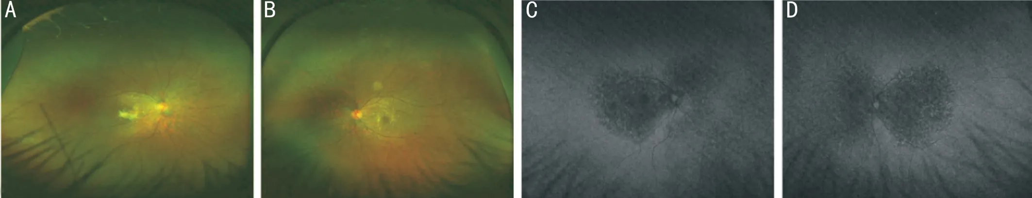
AF images help indicate the extent of RPE-choriocapillaris atrophy and photoreceptor cells loss in BCD. In the regions of RPE-choriocapillaris atrophy, the autofluorescence is decreased. The hypo-fluorescence region extends beyond the posterior pole with the disease’s progress until there is no fluorescence in the posterior pole in the advanced stage.Hyper-fluorescence spots scatter around the hypo-fluorescence regions in the posterior pole. Though autofluorescence in early stage is usually normal, hyper-fluorescence can sometimes be observed
.
Early symptoms of BCD are similar to other autosomal recessive RP, including gradual reduction of visual acuity,paracentral scotomas, visual field constriction, night blindness, and color vision defects. Severe cases may progress to legal blindness at late stages about 50-60s
. Phenotype polymorphism has been observed, sometimes patients from the same family with the same mutations can have different phenotypes.Choroidal neovascularization (CNV) is associated with RPE and Bruch’s membrane breaches. It is rare in hereditary retinal degeneration, except for a few conditions, including BCD. As a complication of BCD, it could be a severe threat to vision.There has been no previous report of any mutations associated with CNV in BCD.
對于剛剛走進大學(xué)人文社會學(xué)科的學(xué)生來說,首要任務(wù)是學(xué)習(xí)專業(yè)知識,接受已有人類文化成果。但這不是我們的最終目的,最終目的是在掌握已有人類文化成果的前提下,進一步走向哲學(xué)社會科學(xué)前沿,承擔思想理論創(chuàng)新發(fā)展的任務(wù)。今天講的這個題目,對于剛上大學(xué)的學(xué)生而言,似乎很遙遠,但不是遙不可及的。在你們經(jīng)過幾年本科、幾年研究生學(xué)習(xí)之后,將會越來越趨近這個目標。立志不怕年少,越早立下志向,越有利于未來的發(fā)展。在我們的學(xué)術(shù)道路上,有兩個“前”目標即前提和前沿,是有志于思想理論創(chuàng)新的學(xué)者必須面對和解決的問題。
The discoveries of the
mutations reported before allowed ophthalmologists to test the correlation between genotype and phenotype. In this study, we report the results of clinical examinations of a Chinese BCD patient and genetic analyses of the
gene of her family. We identified three
mutations in mutation screening of the
gene, one of which was novel.
路基基層表面狀況直接影響防水層的施工質(zhì)量,必須按照基層表面要求對基層進行檢查。基本要求是:基層表面必須平整、干凈和干燥。主要的基層表面處理事項是:用鋼絲或噴砂法處理掉路基表面的浮漿,用蘇打水或燒堿清除掉表面油污,用鬃刷或水清掃掉表面的灰塵、石屑及沙粒等殘留物質(zhì),用打磨機對路基表面凸起進行研磨,用瀝青砂或水泥漿填充路基表面凹陷,從而保證基層表面的平整干凈。
SUBJECTS AND METHODS
This study was conducted at the Department of Ophthalmology, Peking University People’s Hospital, Beijing, China. The study was performed under the Declaration of Helsinki tenets after approval by Ethics Committee of Peking University People’s Hospital(2020PHB250-1). Informed consent was obtained from all individuals included in the study.
A 30-year-old female (the daughter of the family) with suspected BCD and her family all underwent ophthalmologic examinations, including best-corrected visual acuity (BCVA),intraocular pressure (IOP), slit-lamp examination, dilated fundoscopy in order to exclude other ocular diseases. The proband patient underwent comprehensive ophthalmic examinations, including fundus autofluorescence (AF), fundus photography (FP), fundus fluorescein angiography (FFA),Ganzfeld full-field electroretinograms (ERGs), Humphrey visual field, optical coherence tomography (OCT), and optical coherence tomography angiography (OCTA). Clinical diagnosis was made based on tiny sparkling yellowish crystals in the posterior pole, and the atrophy of RPE and choriocapillaris.
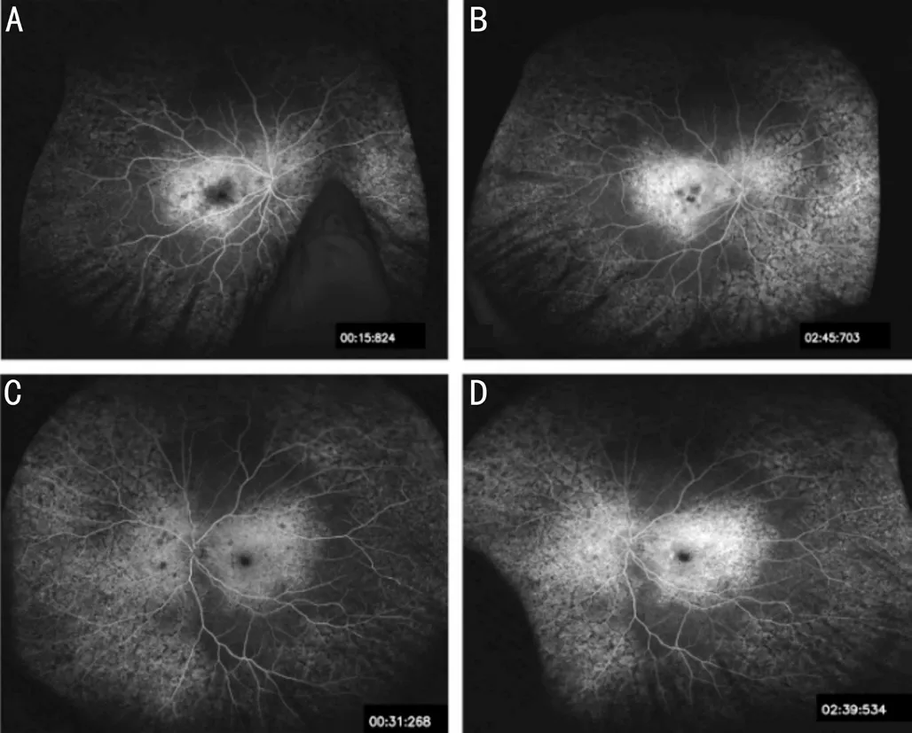
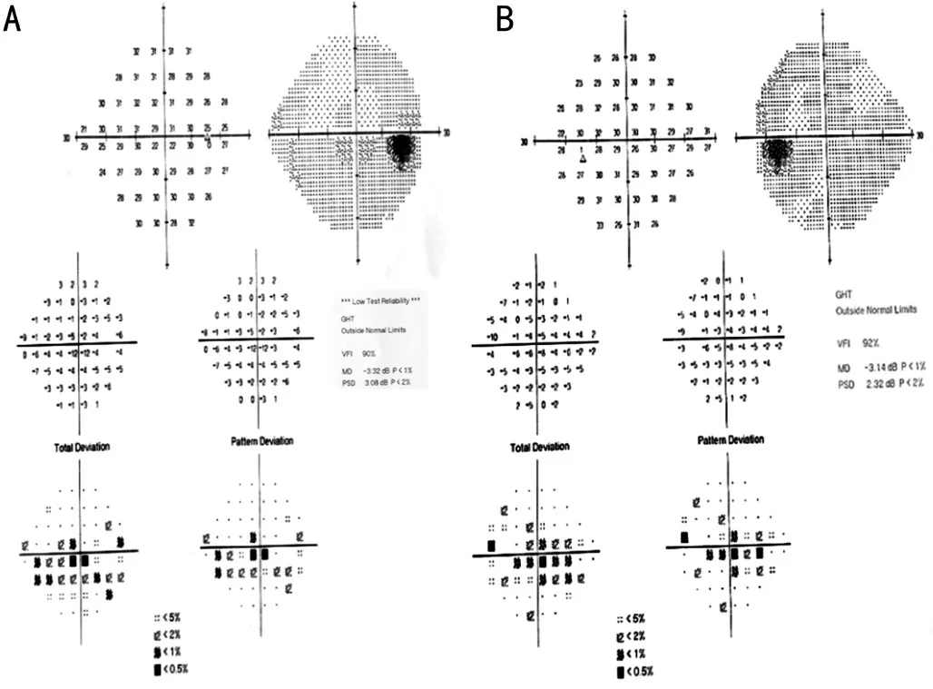
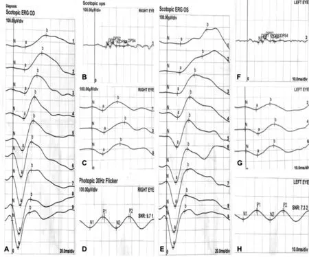
Her BCVA was 20/100 (0.20 logMAR) of right eye, and 20/25(0.8 logMAR) of left eye. Her IOP was normal. Slit lamp examination showed no remarkable anterior segment findings.No crystalline deposits in the cornea and corneal limbus have been observed. Fundus examination showed glittering yellowish crystalline deposits in the posterior pole with diffuse RPE atrophy (Figure 1). AF images showed diffuse hypofluorescence interspersed with hyper-fluorescence scatters in and beyond the posterior pole (Figure 1). OCT images showed thinning and loss of the RPE layer, interruption and loss of the interdigitation zone (IZ) and ellipsoid zone (EZ). Residual EZ layer and RPE can be observed at the fovea. Outer retinal tubulations (ORTs) and atrophy of the choriocapillaris has been observed. OCT and OCTA also demonstrated that active type 2 choroidal neovascular membrane in both eyes (Figure 2).The FFA images showed widespread window defects with multiple scattered hypo-fluorescent spots and leakage in the fovea indicating active CNV in both eyes (Figure 3). Visual field revealed central scotoma and paracentral scotoma in both eyes (Figure 4). Full-field ERG showed a slightly decreased b-wave amplitude in scotopic ERG (Figure 5). Ophthalmic examinations of other family members showed no remarkable findings.
電光源顯微鏡的好處是借助于底座強烈的光線把標本照亮,經(jīng)適當?shù)恼{(diào)節(jié)可以清楚看到物像,但有時會出現(xiàn)故障。光源燈不亮原因在于:(1)電源沒接通;(2)插座插頭未插好;(3)開關(guān)未開;(4)調(diào)節(jié)閥未開;(5)燈泡損壞;(6)保險絲損壞等。
The early changes of the visual field are mainly central or paracentral scotomas. As the disease progresses, the visual field can be constricted or progress to tubular vision.Electrophysiological tests such as full-field ERG and multifocal ERG can help estimate the severity of degeneration of retinal function. Full-field ERG can reflect dysfunction of rod and cone, varying from normal to amplitudes reduction,even undetectable, depending on disease severity
. But sometimes, full-field ERG can remain normal even in the advanced stages of BCD
. Multifocal ERG can accurately identify the retina’s dysfunctional areas, especially when fullfield ERG shows normal results. As the disease progresses,electrooculography (EOG) may show a decrease in the Arden ratio, even earlier than ERG changes
.
RESULTS
The proband, a 30-year-old female, presented with decreased vision with metamorphopsia in her right eye for half a year.The patient received bilateral refractive surgery in 2013 before which she had myopia of -6.00 diopter of spherical power (DS)in both eyes. The patient’s medical records showed she used to attain normal visual acuity in both eyes post refractive surgery.There was no other history of diagnosed eye conditions.
The genomic DNA of four members in this family was extracted from their peripheral blood with a High Pure PCR Template Preparation Kit (Roche, Basel, CH, USA). DNA fragments were enriched by solution-based hybridization capture, then sequenced on the Illumina Miseq platform(Illumina, San Diego, CA, USA) with the 2×300-bp pairedend read module. The hybridization capture procedure was performed with SureSelect Library Prep Kit (Agilent, Santa Clara, CA, USA). Deep intronic sequences were not targeted to avoid decreasing the mean sequence coverage. DNA was sheared
sonication with Diangenode Bioruptor
Plus and hybridized with biotinylated RNA oligonucleotide baits.Captured fragments were removed by using streptavidincoated magnetic beads (Dynabeads
MyOne? Streptavidin T1, Thermo Fisher Scientific) and then eluted. The enriched fragment library was amplified by PCR using primers specific to the linked Illumina adaptors. The resulting libraries were quantified using Q-PCR before proceeding to the Illumina Miseq platform. All samples were sequenced together and 6-bp index sequences (Illumina) were used to distinguish samples.Raw reads from samples were sorted by index sequences after sequencing. Adapter sequences were trimmed by Cutadapt.SolexaQA was used to remove low-quality bases. Clean Reads were aligned to the human reference genome (hg19)with Burrows-Wheeler Aligner (BWA; v.0.7.11). Realignment around known indel sites and Base Quality Score Recalibration(BQSR) were performed by GATK (v.3.3). Human Gene Mutation Database, dbSNP138, 1000 Genome project,ClinicVar, and Exome Sequencing Project were used to screen variants. To detect copy number variants, the sequencing depth of each region covered by probes was calculated according to the alignment files. Functional effect prediction was evaluated by Provean, PolyPhen-2, and Mutation Taster. After candidate causative mutations were determined, the samples were sequenced by Sanger sequencing to verify the mutations and performed with genotype-phenotype co-segregation analysis.

Compared with the complete sequence of the coding and adjacent intron regions of the
gene, three heterozygous mutations were found in the proband patient: c.802_807del(p.V268_I269del), c.810delT (p.E271Nfs*5) and c.1388G>A(p.G463D). The father had c.802_807del mutation and c.810delT mutation. The mother had c.1388G>A mutation.The brother had no mutation (Table 1).
The substitution mutation c.1388G>A is a novel mutation never been reported in the Human Gene Mutation Database(HGMD) and predicted to be pathogenic. The score values of the novel mutation based on three bioinformatics tools are high, suggesting that the mutation causes protein dysfunction and is harmful to the patients. The c.810delT and c.802_807del are deletion mutations that have been reported in BCD patients as pathogenic mutations
.

DISCUSSION
Since the mutation of the
gene was first reported,the pathogenic mechanism of BCD has been intensively studied, and it has been described as a disorder related to lipid metabolism. The CYP4V2 protein is expressed in almost all kinds of tissues and cells, higher in retinal and RPE cells while relatively lower in corneal cells
. As an enzyme, the CYP4V2
protein is responsible for the oxidation of substrates during fatty acid metabolism, especially ω-hydroxylation reactions of medium-chain saturated fatty acids. In BCD patients, the conversion of fatty acid (FA) precursors into n-3 polyunsaturated fatty acid (PUFA) is lower than normal, such as the conversion of eicosapentaenoic acid (EPA, 20:5 n-3) and docosahexaenoic acid (DHA, 22:6 n-3) from α-linolenic acid(18:3 n-3)
. The eye is rich in DHA, and the photoreceptor cells are the richest in all cells
. The membrane disks in the outer segments of photoreceptor cells contain rhodopsin,phosphatidylcholine, and phosphatidylethanolamine, which converted to DHA and other FA
. RPE cells have efficient mechanisms for DHA utilization and recycling, so the cycle of lipids between the outer segments of rods and RPE cells is greatly dependent on the normal functions of RPE cells. This cycle is essential for maintaining normal visual functions so that RPE cells may play an important role in BCD
. Zhang
have demonstrated that PUFA played an important role in mitochondrial damage inducing RPE degeneration in BCD patients and suggested that adeno-associated virus 2(AAV2)-mediated gene therapy may be used as the treatment of BCD. Furthermore, some FA metabolites are involved in anti-inflammation, immunoregulation and so on, so that the defections of CYP4V2 protein function may influence the signaling pathway and even influence other aspects
.Therefore, the mutations of the
gene may have broad influences, and we still need more detailed studies to clarify the mechanism of BCD.
The novel mutation c.1388G>A may be a possible cause that could induce the clinical phenotype of BCD.
The OCT images can reveal the location of crystals and the abnormal changes in the choroid and retinal layers. As reported,the crystals are usually located in the RPE-Bruch’s membrane complex. They can also be located in the neuroepithelial layer retina, the posterior hyaloid membrane, the space between the retina and vitreum
. The disorder of the outer retinal layer in BCD, such as the thinning and disruption of RPE, the loss of IZ, and the interruption of EZ, is progressively severe as natural history of the disease. ORTs are considered only appearing in the regions RPE damaged due to the disruption and rearrangement of photoreceptor cells
. ORTs are more frequently seen in BCD than other degenerative retinal diseases such as age-related macular degeneration (AMD)and RP. The presence of ORTs indicates damaged function of photoreceptor cells and a poor visual prognosis
. OCT images can also indicate the atrophic change of the choroid.Furthermore, central macular thickness (CMT) measure on OCT can be useful in assessing visual prognosis
.
BCD is caused by mutations of the
gene (Gene ID 285440, OMIM 210370), with multiple genotypic and phenotypic variations
. The
gene is located on chromosome 4 (4q35), comprising of 11 exons and encoding a protein of 525 amino acids, a member of the cytochrome P450 protein family
. The protein serves as an enzyme in fatty acids metabolism.
高職院校學(xué)生對學(xué)生事務(wù)管理滿意度的影響因素次序分析……
登錄APP查看全文
猜你喜歡
當代陜西(2022年6期)2022-04-19 12:12:20
當代陜西(2021年8期)2021-07-21 08:31:42
作文大王·笑話大王(2021年4期)2021-04-26 19:00:35
人大建設(shè)(2020年2期)2020-07-27 02:47:54
當代陜西(2019年6期)2019-11-17 04:27:38
當代陜西(2019年13期)2019-08-20 03:54:10
電影(2018年9期)2018-11-14 06:57:21
作文世界(小學(xué)版)(2018年4期)2018-10-16 17:13:34
快樂作文·低年級(2016年12期)2017-01-03 20:52:44
快樂作文·低年級(2016年6期)2016-06-24 18:58:40
 International Journal of Ophthalmology
2022年6期
International Journal of Ophthalmology
2022年6期
- International Journal of Ophthalmology的其它文章
- Intraocular lens removal or not during vitrectomy for acute infectious endophthalmitis after cataract surgery
- Vitreous function and intervention of it with vitrectomy and other modalities
- Short-term outcomes of mitomycin C-augmented excisional bleb revision with capsulectomy for failed Ahmed glaucoma valve
- Evaluation of nintedanib as a new postoperative antiscarring agent in experimental extraocular muscle surgery
- Multimodal imaging of experimental choroidal neovascularization
- A novel Nance-Horan syndrome mutation identified by next-generation sequencing in a Chinese family