Influence of matrigel on the shape and dynamics of cancer cells
2019-11-06 00:46:56TengYe葉騰andFengQiu邱峰
Chinese Physics B 2019年10期
Teng Ye(葉騰) and Feng Qiu(邱峰),2,?
1First Clinical Medical College,Nanchang University,Nanchang 330006,China
2Department of Oncology,First Affiliated Hospital of Nanchang University,Nanchang 330006,China
Keywords:cancer cell,matrigel,migration,shape deformation
1.Introduction
Most kinds of malignant tumor cells are motional and metastatic.They are able to migrate to other tissues and form multiple distant lesions in the late stage.Multiple tumor lesions release cachexia,severely consume body nutrients,and finally cause the death. Studying the mechanisms of cancer cell invasion and metastasis contributes to finding ways to prevent cancer metastasis and cure them at the primary site.It is generally accepted that the tumor microenvironment plays an indispensable role in the tumor invasion and metastasis.The tumor microenvironment contains several kinds of stromal cells(including endothelial cells,[1]tumor-associated fibroblasts,[2]infiltrating immune cells[3]),extracellular matrix(ECM),cytokines,growth factors,inhibitors,nutrients,and oxygen.[4]Due to the existence of the tumor cells,the components are different from normal tissues. For instance,the rapid growth of a tumor results in a hypoxia microenvironment,which conversely influences the proliferation,invasion,migration,and clinical drug resistance of the tumor cells.[5]In addition,when the interaction between normal tissues and ECM becomes awry,cancer occurs in an extended time.[6]So the interaction between tumor cells and the microenvironment could be the key point to regulate the tumor behaviors.[7–9]
Extracellular matrix,which is a fiber macromolecular network,is considered to be the main component of the tumor microenvironment.Previous researches about breast cancer in mice models indicated that ECM plays a crucial role in tumor invasiveness and formation.[10]Also,breast cancer tumorigenesis was found to be relevant with ECM stiffening,collagen crosslinking,and focal adhesion.[11]Basement membrane(BM)is a specific form of ECM,which is composed of collagen type IV,laminins,nidogen,and sulfated proteoglycans.BM lays under epithelial and endothelial cells,separating them from the stromal tissue.[12,13]In the process of tumor metastasis,tumor cells first degrade the epithelial BM and invade into the stromal tissue,before migrating to distant organs or tissues by blood circulation.[12]
Matrigel is a BM extract derived from mice EHS tumor cells.[14]Matrigel functionally influences the growth,invasion,and morphological changes of tumor cells. It can be applied in an in vitro experimental model to mimic the in vivo microenvironment.[15]At the molecular level,Prince et al.studied the matrigel influence on colon cancer microRNAs level,suggesting that matrigel alters cell adhesion,proliferation,and invasion-related mRNAs expression and thus modulates the behaviors of the tumor cells.[16]Recent researches demonstrated that matrigel improves the growth environment of single and multiple tumor spheroids in a three-dimensional(3D)model.[17]However,little is known about the influence of matrigel on individual tumor cells.In this study,we investigate how external matrigel may influence the shape and dynamics of breast cancer cells(MDA-MB-231-GFP cells).Our results show that matrigel facilitates cancer cells’migration and shape deformation.
2.Materials and methods
2.1.Cell line and culture
MDA-MB-231-GFP cells were obtained from Robert Gillies(H.Lee Moffitt Cancer Center,Tampa,FL,USA).The cells were cultured in a medium consisting of Dulbecco’s modified Eagle’s medium(DMEM,Corning),10%fetal bovine serum(FBS,Gibco),100 units/ml penicillin(Corning),and 100μg/ml streptomycin(Corning).They were cultured in an incubator with 5%CO2at 37?C.
2.2.Sample preparation and microscopy tracking
We developed an experimental model to study how cells behave under matrigel and drugs[Fig.1(a)].The cells were trypsinised with Trypsin-EDTA(Corning),centrifuged,and re-suspended by using the culture medium. Then,the cells were counted under an optimal microscope(TS-100,Nikon)by using a cell-count board and seeded into a 96-well plate with a density of 1000 cells per well.After an over-night incubation,the adherent cells were covered with matrigel matrix(Corning BioCoat,standard version),and incubated at 37?C for 30 min to solidify the matrigel.Then,200μl culture medium was added onto the matrigel.In the control experiments without matrigel,we directly added 200μl culture medium on the adherent cells after an over-night incubation.The matrigel concentration can be varied by mixing the culture medium with pure matrigel. After the sample was finished,an inverted microscope(Ti-ECLIPSE,Nikon)was used to track the MDA-MB-231-GFP cells under fluorescence field for 8 hours with a×10 objective lens.The time interval between each two images was 5 min.
2.3.Image processing and analysis
The fluorescent images were used to analyze the mean squared displacement(MSD),speed,area,and shape factor of the cells.For the MSD calculation,each cell trajectory was tracked semi-automatically by using the CellTracker plugin in Matlab 2014a.[18]The calculated coordinates were used to plot the curve of MSD by a macro command.[19]Only single cells were selected to calculate the MSD.A MSD curve of 5 hours was plotted from 8-hours’tracking data by using the slidingwindow averaging method.Each 5-hour curve approximately contained the tracking data of 200 cells.
To obtain the cells’areas and shape factors,ImageJ was applied to calculate the tracked fluorescent image stack.The calculation was based on the brightness of the cells under fluorescent field.We first converted the 16-bit fluorescent image stack into an 8-bit one and used the function”Analyze Particles”to calculate single cell areas(between 300μm2and 2500μm2).Meanwhile,the cells’perimeters were also calculated.Given the area and perimeter,we could then obtain the shape factor,which is defined as the ratio between the perimeter and the square root of the area. The number of cells in each group we used was 4000.For the groups under different conditions,we calculated,respectively,the average and the distribution of the area and the shape factor.
3.Results
We tracked fluorescent images under a Ti-ECLIPSE microscope[see Fig.1(a)]and analyzed the trajectories of the cells under different conditions by CellTracker plugin in Matlab 2014a.Figure 1(b)shows the observations of two typical cells under different conditions,from which we can clearly see the cell placed under 100%matrigel has a larger spreading area and longer motion track.
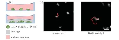
Fig.1. (a)Schematic of the experimental setup,and(b)typical experimental observations under different conditions,where the red lines represent the trajectories of the selected cells.
3.1.Cells show higher migration persistence and speed under matrigel
We now investigate quantitatively how matrigel influences the cell shape and migration.We start with the migration and focus on two quantities:MSD and speed.As shown in Fig.2(a),all the MSD curves increase approximately linearly with time,indicating that the cells perform Brownian-motionlike random walks;the MSD curves of the groups with matrigel increase with time much faster than that of the group without,and the one with 100%matrigel slightly faster than that with 50%matrigel.When there is no matrigel,most cells have a speed slower than 1μm/min[Fig.2(b)];and the number of cells with a speed higher than 1μm/min increases dramatically when matrigel is planted on top[Figs.2(c)and 2(d)];in addition,the probability for the cells to have a speed in 0–0.3μm/min is reduced as the concentration of matrigel is increased from 50%to 100%[Figs.2(c)and 2(d)].These results indicate that matrigel facilitates cell motion:it enhances not only the speed but also the migration persistence of cancer cell migration.
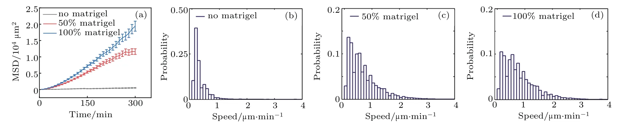
Fig.2.(a)MSD of the cells under different conditions,and(b)–(d)respective speed distributions.
3.2.Cells are more spread and elongated under matrigel
We now investigate the shape change of the cells under different conditions.We first measure the 2-dimensional area of the cells.As shown in Fig.3(a),when covered with matrigel,the cell’s average area is 978.44μm2(100%matrigel)and 976.85μm2(50%matrigel),respectively,while that without matrigel is only 757.91μm2[Fig.3(a)];when there is no matrigel,the peak area is 400–600μm2[Fig.3(b)],and it increases to 700–1200μm2when matrigel is planted[Figs.3(c)and 3(d)].The enhancement in the cell area suggests a stronger adhesion of the cells to the matrigel and/or the bottom of the plate.
We proceed to study the shape factor,which is defined as the ratio between the perimeter and the square root of the area of the cell images.The shape factor is a quantity widely used to measure cells’shape deformation.[20]As shown in Fig.3(e),the average of the shape factors is obviously increased when matrigel is added.From Fig.3(f),we can see that the shape factors for the case of no matrigel mostly locate between 3.7 and 4,which are close to the value of a circle’s shape factor(~3.54);when the cells are covered with matrigel,most cells’shape factors are distributed in the interval of 4–6[Figs.3(g)and 3(h)].The differences between the results of 100%matrigel and those of 50%matrigel are negligible.These results indicate that matrigel facilitates cells’shape deformation and elongation.

Fig.3.(a)Area average and(b)–(d)corresponding area distributions;(e)shape factor average and(f)–(h)corresponding distributions.
4.Conclusion
In summary,we experimentally investigated the influence of matrigel on the shape and dynamics of breast cancer cells(MDA-MB-231-GFP cells)and obtained that the cells tend to elongate,become larger,and migrate faster in the presence of matrigel.The differences between the influence of 100%matrigel and that of 50%matrigel are however negligible.Our results show explicitly the correlation between the shape and migration speed of the cancer cells when the influence of matrigel is taken into account.The detailed mechanism underlying these influences and the correlation will be investigated in near future.
Acknowledgments
The authors thank the support of the CAS Key Lab of Soft Matter Physics,where part of this work was done,and thank Xiaochen Wang for very helpful discussions.

- Chinese Physics B的其它文章
- Theoretical analyses of stock correlations affected by subprime crisis and total assets:Network properties and corresponding physical mechanisms?
- Benefit community promotes evolution of cooperation in prisoners’dilemma game?
- Theory and method of dual-energy x-ray grating phase-contrast imaging?
- Quantitative heterogeneity and subgroup classification based on motility of breast cancer cells?
- Designing of spin filter devices based on zigzag zinc oxide nanoribbon modified by edge defect?
- Opto-electromechanically induced transparency in a hybrid opto-electromechanical system?