Value of superb micro-vascular imaging in predicting ischemic stroke in patients with carotid atherosclerotic plaques
2019-04-16 08:20:58DeBinYangJieZhouLanFengRongXuYingChunWang
World Journal of Clinical Cases 2019年7期
De-Bin Yang,Jie Zhou,Lan Feng,Rong Xu,Ying-Chun Wang
Abstract
Key words: Superb micro-vascular imaging;Contrast-enhanced ultrasound;Carotid atherosclerotic plaques;Ischemic stroke
INTRODUCTION
Effective preventive measures are important ways to prevent ischemic stroke[1,2].Studies have revealed that carotid atherosclerotic plaque is an independent risk factor for ischemic stroke[3,4].Carotid plaques cause stenosis of the lumen,and unstable plaques are also prone to rupture and to form thrombi[5,6].Thrombosis can block distal intracranial blood vessels leading to ischemic stroke[7,8].Researchers have found that neovascularization in the plaque is closely related to the stability of a plaque[9].Contrast-enhanced ultrasound (CEUS) can sensitively detect neovascularization in the plaque and then determine the stability of the plaque.It is currently the main clinical detection method for assessing plaque stability[10,11].However,patients must be injected with contrast agents to perform CEUS,which requires the assistance of a nurse.The operation is more complicated than conventional ultrasound examination and requires more examination time.Therefore,it cannot be a method for screening unstable plaques.With the development of new ultrasound technology,the emergence of superb micro-vascular imaging (SMI) provides a more sensitive method in detecting low-velocity blood flow than color Doppler,providing the possibility of clinical screening for unstable plaques[12,13].Therefore,the present study compared the accuracy of SMI and CEUS in predicting ischemic stroke and explored the potential value of SMI in predicting ischemic stroke.
MATERIALS AND METHODS
Study object
Carotid atherosclerosis patients with a lumen area stenosis of 50%-70% who were confirmed by conventional X-ray angiography in Jiading District Central Hospital Affiliated Shanghai University of Medicine &Health Sciences from March 2013 to March 2015 were recruited.These patients underwent carotid ultrasound and at least one side of carotid atherosclerotic plaque was confirmed.In total,130 patients with complete clinical data were included in the study.Patients with hemorrhagic stroke,brain trauma,poisoning,post-epileptic state,hypertensive encephalopathy,abnormal blood glucose,encephalitis and vital organ function were excluded.All patients provided informed consent,and the study was supported by the Ethics Committee of Jiading District Central Hospital Affiliated Shanghai University of Medicine &Health Sciences.
Research methods
Clinical data records:The clinical data of each patient,including age,gender,blood lipid index (cholesterol,triglyceride,high-density lipoprotein,low-density lipoprotein,lipoprotein a),smoking history,diabetes history,hypertension history,and high-sensitivity C-reactive protein,were recorded.
Ultrasound examination:Toshiba Aplio500 ultrasound (PLT-1005BT linear array probe,5-10 MHz) equipped with SMI imaging software was used in the present study.GE Logiq E8 ultrasound (9L4 probe,4-9 MHz) equipped with ultrasound contrast imaging was also used in the present study.The patient was placed in a supine position with the head biased to the opposite side,and the neck was fully exposed.The carotid artery was continuously scanned longitudinally and laterally to observe the presence of plaque and plaque echo.The position,length and thickness of the target plaque were recorded.If the patient had multiple plaques,then the thickest hypoechoic or mixed echo plaques were selected as the target plaque.
SMI examination:After clearly displaying the two-dimensional ultrasound image of the plaque,the patient was instructed to continue to breathe calmly.The target plaque was observed using the transverse and longitudinal sections of the SMI imaging mode.The video of the target plaque was recorded.After the scan was finished,the video was played back and was observed whether there were new blood vessels in the plaque.The blood vessels in the plaque were categorized as follows[14]:level 0:no blood flow signal is found in the plaque;level I:one or several dot blood vessels were found in the plaque;level II:dot blood vessels and 1-2 linear blood vessels are found;and level III:Multiple linear blood vessels are visible in the plaque,and most of them penetrate the plaque.
CEUS examination:Sonovue microbubble contrast agent (Bracco,Italy) and 5 mL of physiological saline were used to prepare a suspension.The truncated median vein was punctured and injected with 2.0 mL of contrast agent.After the injection,5 mL of physiological saline was injected immediately,and the contrast mode was started.The enhancement of the plaque was observed,and the video was retained for later analysis.The contrast video was played back and the contrast perfusion of the plaque was observed.The image was analyzed by contrast analysis software,and the region of interest was manually drawn.The time-intensity curve was plotted to obtain the contrast parameters:time-to-peak and plaque enhancement intensity (EI)[15,16].
Patient follow-up and grouping
A total of 130 patients who participated in the study were followed up for 3 years.The first follow-up was performed at 3 mo after the end of treatment.Subsequently,follow-up was performed every 6 mo by phone.The endpoint was defined as ischemic stroke occurring during patient follow-up.Patients’ refusal,midway exit,and accidental death were defined as loss of follow-up.Based on the follow-up results,patients with endpoint events were defined as the stroke group and the remaining patients were defined as the non-stroke groups.The baseline data,imaging parameters,blood lipids (cholesterol,triglyceride,high-density lipoprotein,lowdensity lipoprotein,lipoprotein a),high-sensitivity C-reactive protein,smoking history,diabetes history,hypertension history,SMI and CEUS diagnostic parameters were analyzed.
Statistical analysis
Statistical analysis was performed using Statistical Product and Service Solutions and Medcalc software.The measurement data was expressed as x ± s,and the count data was expressed in frequency.The comparison between the two groups of measurement data was performed using an independent samplettest,the count data was applied by chi-square test,and the graded data was performed using Subparameter test.Correlation analysis between SMI level and EI was performed using Spearman correlation analysis.Multivariate Cox proportional regression was used to analyze the risk of stroke in patients.The receiver operating characteristic (ROC) curve was used to evaluate the accuracy of the potential indicators in predicting ischemic stroke.The incidence of ischemic stroke in patients with different SMI levels was analyzed by Kaplan-Meier,and the difference was analyzed by log-rank.P< 0.05 was considered statistically significant.
RESULTS
General data analysis of patients
By the end of the follow-up,five patients were lost to follow-up and forty-three patients were diagnosed with ischemic stroke and placed in the stroke group.There were 82 patients in the non-stroke group.The general information for the two groups of patients was presented in Table 1.Comparison of the clinical data between the two groups found that the age,gender,plaque thickness,lipoprotein a,high-density lipoprotein,high-sensitivity C-reactive protein,cholesterol,triglyceride,low-density lipoprotein,smoking history,diabetes history and hypertension history in the two groups were similar.There was no significant difference between the two groups (P>0.05).
The number of SMI vascular grading cases in the stroke and non-stroke groups were as follows:level 0:1 case and 17 cases,respectively (Figure 1A);level I:6 cases and 48 cases,respectively (Figure 1B);level II:17 cases and 14 cases,respectively(Figure 1C);and level III:19 cases and 3 cases,respectively (Figure 1D).Patients in the stroke group were mainly in level II and III,while patients in the non-stroke group were mainly in level 0 and I.There were statistically significant differences between the two groups (Z= 56.678,P< 0.001).
There was no statistically significant difference in plaque time-to-peak between the stroke group and the non-stroke group (t= 1.618,P= 0.108).EI in the stroke group(Figure 2A) was larger than that in the non-stroke group (Figure 2B),and the difference was statistically significant (t= 7.526,P= 0.000).
Multivariate COX regression analysis of ischemic stroke
Multivariate analysis of SMI level and EI showed that SMI level (P= 0.013) and EI (P= 0.032) were the independent influencing factors affecting ischemic stroke (Table 2).
Correlation analysis of SMI level and EI
Spearman correlation analysis revealed a positive correlation between SMI level and EI (r= 0.737,P= 0.000;Figure 3).
Diagnostic efficacy of SMI grade and EI in predicting ischemic stroke in patients with plaque
According to the ROC analysis,the area under curve (AUC) of SMI level predicting ischemic stroke in patients with plaque was 0.878.The best diagnostic point was ≥level II.The sensitivity was 86.05%,and the specificity was 79.27%.The AUC of EI predicting ischemic stroke in patients with plaque was 0.890.The best diagnostic point was 9.92 db.The sensitivity was 88.37%,and the specificity was 89.02%.The difference was similar and there was no statistical significance between them (Z=0.336,P= 0.737;Figure 4).
Survival curve analysis of patients with ischemic stroke under different SMI classification
Survival curve analysis of patients with ischemic stroke under different SMI classification was plotted by Kaplan-Meier.The results showed that with the increase of SMI level,the incidence of ischemic stroke was gradually increased.The log-rank test revealed a statistically significant difference (X2= 108.931,P= 0.000).The incidence rate of SMI level 0 and level I was similar.The incidence rate of SMI level II was significantly higher than level I.Therefore,it is feasible to use the SMI level II as a predictor of ischemic stroke (Figure 5).
DISCUSSION
Carotid atherosclerotic plaques are prone to cause ischemic stroke.Studies have shown that plaque formation is an independent risk factor for stroke[17,18].Plaques gradually block blood vessels and cause stroke as the disease progresses.In addition,unstable plaques are prone to rupture,resulting in the blockage of distal blood vessels and subsequent ischemic stroke.Studies have found that neovascularization in plaques is an important indicator of plaque stability[19].Therefore,timely detection and effective assessment of neovascularization in plaques have potential value in predicting patients with ischemic stroke.Recent studies have shown that CEUS is of great value in the detection and assessment of neovascularization in plaques[20,21].The operation of CEUS is complex and requires the support of contrast agents and nurses.Therefore,it cannot be used as a screening tool for finding unstable plaques.SMI is a novel technique for detecting blood flow in micro-vessels,enabling the detection of micro-vessels at a much lower speed[13,22-24].However,there are few studies on the assessment of neovascularization in plaque,and it is not clear whether it can be used to predict ischemic stroke in patients with carotid plaque.Therefore,the present study aimed to determine the ability of SMI in detecting neovascularization of carotid atherosclerotic plaque and analyzed its value in predicting ischemic stroke in patients with plaques.

Table 1 Comparison of clinical baseline data between the two groups,n (%)
The subjects recruited in this study needed to meet two conditions.First,the degree of vascular stenosis needed to be between 50% and 70%.Second,the patients were only treated with conservative drugs in order to prevent ischemic stroke caused by excessive plaque growth.These conditions controlled to the best of our ability that an ischemic stroke in the study subject was caused by the rupture of an unstable plaque.
In this study,baseline data analysis was performed on patients who underwent 3 years of follow-up.The results showed that the clinical data of age,gender,serum index,and medical history were not significantly different between the two groups (P>0.05).It indicates that the difference in patient baseline data is small.However,SMI level and EI were different between the two groups.The results showed that patients in the stroke group had higher SMI grades and higher EI values.Multivariate COX regression analysis found that SMI level and EI were independent factors influencing ischemic stroke.This initially suggests that SMI levels and CEUS markers can be used to detect neovascularization in plaques of patients with carotid atherosclerosis and to assess the occurrence of ischemic stroke.Previous studies have reported that unstable plaques are prone to rupture and cause distal vascular obstruction to cause ischemic stroke.The abundant neovascularization in the plaque is associated with rupture of the plaque[25,26].Therefore,SMI and CEUS are good tools for dynamic observation of blood flow distribution in the plaque,which can be used to assess the occurrence of ischemic stroke .
Studies have shown that contrast EI in plaques have a good correlation with histological micro-vessel density[27].It has been clinically established that CEUS can be used to analyze the stability of plaques[20,28].Therefore,we used correlation analysis to evaluate the value of SMI in detecting low-velocity blood flow in plaques.Spearman correlation analysis showed that SMI level was positively correlated with EI,with a correlation of 0.737,suggesting SMI can evaluate blood flow in plaque like CEUS.
In order to investigate the value of CEUS and SMI in predicting ischemic stroke caused by plaque rupture,the ROC curve was used for analysis,and it was found that both could achieve an AUC of approximately 90%.This indicates that both are excellent predictors of stroke.SMI is more convenient and easier to promote than CEUS,suggesting that the clinical use of SMI can be applied for the large-scale screening of unstable plaques.In addition,the ROC curve revealed that the best diagnostic point of SMI was level II.Furthermore,level 0 and I suggested a negative result,while level II and III suggested a positive result.Based on the survival curve of patients,which compared the incidence of ischemic stroke in different SMI levels,it was found that patients with SMI level 0 and I had a similar incidence rate,but the incidence of level II and III significantly increased.This suggests that SMI level II can be used as one of the diagnostic criteria in clinic.
This study has certain limitations.Although we have screened patients with a stenosis degree between 50% and 70% through strict inclusion and exclusion criteria,we still cannot fully guarantee that the reason for ischemic stroke during follow-up was due to plaque rupture.Some patients may have had a stroke due to plaque clogging,resulting in false positive results.To expand our findings,we plan to establish a rabbit carotid plaque model with anatomical results as a standard for plaque rupture to accurately compare the diagnostic efficacy of SMI and CEUS.
In summary,SMI can be used as a non-invasive method for screening unstable plaques.It has a high diagnostic accuracy and can be applied for the clinical diagnosis and prevention of ischemic stroke.

Figure 1 Superb micro-vascular imaging vascular grading in patients with carotid atherosclerosis.
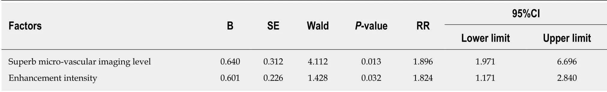
Table 2 Cox multivariate analysis of patients with ischemic stroke

Figure 2 Carotid plaque Tic analyses.

Figure 3 The correlation between superb micro-vascular imaging level and enhancement intensity.

Figure 4 Receiver operating characteristic curve of ischemic stroke in patients with plaques predicted by superb micro-vascular imaging level and enhancement intensity.

Figure 5 Survival curves of patients with ischemic stroke under different superb micro-vascular imaging levels.
ARTICLE HIGHLIGHTS
Research background
Patients with carotid atherosclerotic plaque are prone to ischemic stroke because plaques gradually block blood vessels and cause stroke.Secondly,unstable plaques are prone to rupture and form thrombi,which in turn leads to ischemic stroke.Contrast-enhanced ultrasound (CEUS)can be used to assess unstable plaques because it can detect blood flow in the plaque.However,CEUS is complicated,requires the use of contrast agents and requires the support of nurses,so it cannot be used as a large-scale screening tool for unstable plaques.
Research motivation
Superb micro-vascular imaging (SMI) is a new technique for detecting micro-vascular blood flow,enabling the detection of low-speed micro-vessels.It is expected to be a screening tool for unstable plaques.However,there are few reports on the evaluation of neovascularization in plaques by SMI.Whether it can be used to predict ischemic stroke in patients with carotid plaque is still inconclusive.
Research objectives
In this study,the distribution of micro-vascular blood flow in carotid atherosclerotic plaques was detected by the SMI technique.The aim of this study is to compare the accuracy of SMI and CEUS in predicting ischemic stroke,and to explore the value of SMI in predicting ischemic stroke in patients with unstable carotid plaque.
Research methods
Patients with carotid plaques (luminal stenosis of 50%-70%) were recruited as the subjects in the present study.The clinical baseline data,serological data,CEUS and SMI data were recorded.All patients were followed up for 3 years.During the follow-up period,patients only underwent conservative drug treatment to prevent an ischemic stroke caused by excessive plaque growth within 3 years.According to whether or not ischemic stroke occurred during the follow-up period,the patients were divided into the stroke group and the non-stroke group.The differences in clinical data between the two groups were compared.The correlation between SMI and CEUS was analyzed.The value of SMI and CEUS in predicting ischemic stroke in patients with carotid atherosclerotic plaque within 3 years was explored.
Research results
In the present study,there were 43 patients in the stroke group and 82 patients in the non-stroke group.Patients in the stroke group were mainly in SMI level II and III,while patients in the nonstroke group were mainly in SMI level 0 and I.Cox regression showed that SMI level and enhancement intensity were the independent factors influencing ischemic stroke.SMI level was positively correlated with enhancement intensity.The area under curve of SMI level and enhancement intensity predicting ischemic stroke was 0.878 and 0.890.It indicated that SMI was as good as CEUS in predicting ischemic stroke in patients with unstable carotid plaque.As the SMI level gradually increased,the incidence of ischemic stroke gradually increased.SMI level II revealed the highest accuracy of predicting ischemic stroke in patients with carotid plaque.
Research conclusions
SMI can assess neovascularization in unstable plaques just like CEUS,but SMI is non-invasive and simple to operate.Hence,it can be used as a non-invasive tool for screening for unstable plaques.It can be applied to the clinical diagnosis and prevention of ischemic stroke.
Research perspectives
This study further plans to establish a rabbit carotid atherosclerotic plaque model.The anatomical results of experimental rabbits will be used as criteria for plaque rupture,and the value of SMI and CEUS for predicting unstable plaque rupture will be more accurately analyzed.
 World Journal of Clinical Cases2019年7期
- World Journal of Clinical Cases的其它文章
- Type l congenital extrahepatic portosystemic shunt treated by orthotopic liver transplantation:A case report
- Min-invasive surgical treatment for multiple axis fractures:A case report
- Villous adenoma coexistent with focal well-differentiated adenocarcinoma of female urethral orifice:A case report and review of literature
- Congenital bronchobiliary fistula:A case report and review of the literature
- Robot-assisted gallbladder-preserving hepatectomy for treating S5 hepatoblastoma in a child:A case report and review of the literature
- Non-lnvasive management of invasive cervical resorption associated with periodontal pocket:A case report