3D—ASL結合1H—MRS在常見腦腫瘤術前診斷中的臨床應用研究
2018-01-16 08:44:08陳亞晗柴夢琪李娜陸皓吳為民陸玉敏
右江醫學 2017年6期
陳亞晗+柴夢琪+李娜+陸皓+吳為民+陸玉敏
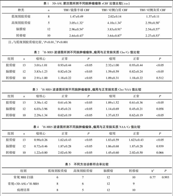
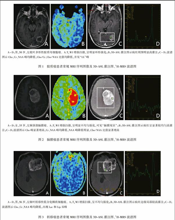
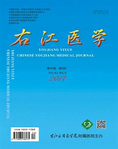
【摘要】目的探討三維動脈自旋標記(3D arterial spin labeling,3D-ASL)結合氫質子磁共振波譜(1H magnetic resonance spectroscopy,1H-MRS)成像在顱腦常見腫瘤診斷中的臨床應用價值。方法選擇經術后病理證實的35例腫瘤病例(其中13例膠質瘤,12例腦膜瘤,10例轉移瘤),測量腫瘤在3D-ASL血流灌注圖中最大腫瘤血流量(tumor blood flow,TBF)與對側正常腦白質、腦灰質、腫瘤鏡像區腦血流量(brain tumor flow,CBF),同時測量波譜圖腫瘤瘤核心及對側正常腦實質感興趣區的膽堿(Cho)、N-乙酰天門冬氨酸(NAA)、肌酸(Cr)峰值及其比值,結合術后病理診斷進行分析。結果高級別膠質瘤與腦膜瘤、轉移瘤灌注值差異無統計學意義(P>0.05),低級別膠質瘤與其他腫瘤灌注值差異有統計學意義(P<0.01);1H-MRS波譜圖顯示膠質瘤、轉移瘤瘤核心、瘤周及腦膜瘤瘤核心的Cho/Cr、NAA/Cr、Cho/NAA代謝物比值大于正常腦實質,腦膜瘤瘤核心Cho/NAA代謝物比值大于其他兩種腫瘤,膠質瘤瘤周代謝物比值大于其他兩種腫瘤。結論3D-ASL與1H-MRS均可作為常規磁共振成像(magnetic resonance imaging,MRI)的重要補充,兩者結合使用對顱腦常見腫瘤的診斷及鑒別診斷有積極臨床應用參考價值。
【關鍵詞】三維動脈自旋標記;氫質子磁共振波譜;腦腫瘤
中圖分類號:R651.1文獻標識碼:ADOI:10.3969/j.issn.1003-1383.2017.06.018
【Abstract】ObjectiveTo explore the clinical value of 3D arterial spin labeling(3D-ASL) combined with 1H magnetic resonance spectroscopy(1H-MRS) in the diagnosis of common brain tumors.Methods35 cases of tumors confirmed by postoperative pathology(13 cases of glioma,12 cases of meningioma,and 10 cases of metastatic tumor)were selected.Tumor blood flow(TBF)in 3D-ASL perfusion images and cerebral blood flow(CBF)in contralateral normal white matter,gray matter and tumor mirror area were measured.At the same time,cholinergic(Cho),N-acetylated aspartate(NAA),creatine(Cr)peak and their ratios in the region of interest of the tumor and the normal brain parenchyma on the contralateral side were measured.Combined with postoperative pathological diagnosis,the above-mentioned situations were analyzed.ResultsThere was no statistically significant difference in the perfusion value of high grade glioma with meningioma and metastatic tumor(P>0.05),while there was statistically significant difference between low grade glioma and other tumor perfusion values(P<0.01).1H-MRS spectroscopy showed that the ratio of Cho/Cr,NAA/Cr and Cho/NAA metabolites in the cores and peritumoral areas of glioma,metastatic tumor and meningioma was greater than that of the normal brain parenchyma,the ratio of Cho/NAA metabolites in the core of meningiomas was significantly greater than that of the other two tumors,and the ratio of metabolites in peritumoral area of gliomas was greater than that of the other two tumors.Conclusion3D-ASL and 1H-MRS can be used as an important supplement to conventional magnetic resonance imaging(MRI).The combination of both of them can be used as reference for the diagnosis and differential diagnosis of common brain tumors.
【Key words】3D-ASL;1H-MRS;brain tumorsendprint