多巴胺受體在大鼠結腸黏膜下神經叢的表達和細胞分布
2017-06-24 11:38:33朱進霞趙文明
首都醫科大學學報 2017年3期
張 悅 李 蘊 朱進霞 趙文明
(1.首都醫科大學基礎醫學院免疫學系,北京 100069;2. 首都醫科大學基礎醫學院生理學與病理生理學系,北京 100069)
· 基礎研究 ·
多巴胺受體在大鼠結腸黏膜下神經叢的表達和細胞分布
張 悅1, 2李 蘊1*朱進霞2趙文明1*
(1.首都醫科大學基礎醫學院免疫學系,北京 100069;2. 首都醫科大學基礎醫學院生理學與病理生理學系,北京 100069)
目的 多巴胺(dopamine, DA)與其受體結合調節腸道動力、黏膜分泌及屏障等,黏膜下神經元參與DA對黏膜的生理調節,但其受體的具體分布尚不明了。通過檢測DA受體在大鼠結腸黏膜下層的蛋白和mRNA表達及細胞分布,為DA調節大鼠結腸病理生理機制提供依據。方法 用RT-PCR和Western blotting方法定性測定DA受體在大鼠結腸黏膜下層的mRNA和蛋白的表達;免疫熒光雙染色后在激光共聚焦顯微鏡下觀察DA受體在黏膜下血管活性腸肽(vasoactive intestinal peptide,VIP)能和膽堿能神經元上的分布。結果 在大鼠結腸黏膜下層,DA受體D1、D2、D5的mRNA及蛋白均有表達;在黏膜下神經叢VIP能陽性的神經元中有大量分布,其中D1陽性的神經元占58.94%±2.245%,D2陽性神經元占52.03%±9.384%,D5陽性神經元占86.21%±2.902%,且D5陽性神經元數量顯著高于D1和D2;在黏膜下神經叢膽堿能陽性的神經元廣泛表達DA受體,其中D1陽性神經元占87.75%±7.307%,D2陽性神經元占88.50%±8.761%,D5陽性神經元占89.25%±10.75%。結論 DA受體在大鼠結腸黏膜下層有D1、D2及D5的表達,且在VIP能及膽堿能神經元上均有分布,其中D5受體在VIP能神經元上的分布顯著高于D1及D2受體。本研究可為深入探討DA調節結腸黏膜生理及病理機制提供形態學依據。
多巴胺;多巴胺受體;黏膜下神經叢;血管活性腸肽;乙酰膽堿
多巴胺(dopamine, DA)作為兒茶酚胺類神經遞質,在中樞及外周均有廣泛分布,其中腸道DA的主要來源為黏膜上皮細胞和腸神經系統[1]。DA受體分有5個亞型D1~D5,均為G蛋白偶聯受體,可分為2類,分別為D1類受體(D1和D5)及D2類受體(D2、D3和D4)。D1類受體主要通過升高胞內的cAMP發揮相應的生理調節作用;D2類受體則通過降低胞內的cAMP來發揮作用。DA與相應受體的結合不僅對胃腸動力具有重要的調節作用[2-3],還對黏膜的分泌及屏障功能發揮重要作用[4]。但因研究方法、腸道節段生理差異及神經因素參與與否等原因,DA發揮功能的受體機制并不一致[5-6],且DA受體在消化道具體分布[7-8]也尚不完全明確。腸神經系統是由上億神經元所形成的腸道神經網絡系統[9],可合成、釋放乙酰膽堿和多種神經肽對胃腸功能發揮重要的獨立整合作用。腸神經系統分為黏膜下神經叢和肌間神經叢,其中黏膜下神經叢主要調節黏膜功能,其中血管活性腸肽(vasoactive intestinal peptide,VIP)和乙酰膽堿(acetylcholine,ACh)是黏膜下促分泌性神經元分泌的主要神經遞質,在黏膜分泌、血供及黏膜免疫等方面發揮重要調節作用[10];肌間神經叢則主要負責調節胃腸動力。研究[11]證明DA可直接作用于腸道肌層或肌間神經元上的相應受體對結腸動力發揮直接或間接的調節作用,但DA對黏膜生理的調節作用及相關受體分布數據相對缺乏。有研究顯示DA對結腸單純黏膜層跨膜離子轉運的調節主要經由腎上腺素能β受體[12],而對結腸黏膜-黏膜下組織的跨膜離子轉運則主要經由D2受體[6],提示黏膜下神經叢可能參與了DA對結腸生理的調節機制,但DA受體在大鼠結腸黏膜下神經叢的分布及具體類型還不明確。本研究旨在探討DA受體在黏膜下神經叢上的表達及細胞定位,為深入探討DA調節黏膜生理功能的機制提供形態學數據。
1 材料與方法
1.1 實驗動物
選取雄性SD大鼠8只(首都醫科大學實驗動物中心提供),動物許可證號:SCXK(京)2012-0001。體質量為200~250 g,經實驗動物福利委員會許可,動物在室溫條件,正常更替光照,24 h食水供應。安樂處死后取結腸組織,其中4只大鼠的結腸用解剖鑷剝離出黏膜下層并凍存于液氮中,另外4只大鼠的結腸腸管于OCT中包埋后經液氮固化后保存于-80 ℃冰箱中。
1.2 RT-PCR檢測DA受體在大鼠結腸黏膜下層mRNA的表達
1.2.1 RNA的提取
取4只適量大鼠遠端結腸黏膜下層組織置于研缽中,加入液氮,用搗錘研磨碾碎組織,加至放有1 mL Trizol(美國Invitrogen公司)的1.5 mL EP管內,輕輕上下顛倒混勻,冰上靜置5 min。加入氯仿0.2 mL,劇烈震蕩15 s,冰上靜置2~3 min,4 ℃低溫、12 000 r/min 高速離心15 min。離心后將EP管傾斜45°取出,將上層水相移入干凈的離心管中,加入異丙醇沉淀RNA(每1 mL Trizol對應0.5 mL異丙醇)。將混合物在室溫條件下孵育10 min,之后4 ℃ 12 000轉高速冷凍離心10 min。移去上層懸液,用75%(體積分數)的乙醇洗滌RNA 沉淀一次(每1 mL的Trizol至少加1 mL的75%乙醇),4 ℃下 7 500 r/min低速冷凍離心5 min,棄上清,簡單干燥RNA沉淀。加入10~20 μL 無酶水溶解RNA樣品,經檢測合格后,進行RNA的反轉錄。
1.2.2 反轉錄
取0.1 ng~5 μg的總RNA,根據RNA溶液的濃度計算出加樣體積,并依次加入1 μL(50 μmol/L)Oligo (dT)和無RNA酶水混合至12 μL,65 ℃加熱5 min后置于冰上,再分別加入如下成分:4 μL 緩沖溶液,2 μL 10 mmol/L dNTP Mix,1 μL RiboLock RNase抑制劑(20 U/μL),1 μL反轉錄酶。將混合物在 42 ℃環境下孵育60 min,以70 ℃孵育5 min終止反應。試劑盒購于美國Thermo公司。
1.2.3 擴增
RT-PCR用于測定大鼠遠端結腸黏膜下層的D1、D2、D3、D4和D55種DA受體mRNA的表達情況。DA受體D1正向引物序列為5′-GGA TGA CAA CTG TGA CAC AAG GTT G-3′,反向引物序列為5′- AAG CTG ATG AGG GAC GAT GAA-3′;DA受體D2正向引物序列為5′- CAC CAC GGC CTA CAT AGC AA-3′,反向引物序列為5′-GGC GTG CCC ATT CTT CTC T-3′;DA受體D3正向引物序列為:5′-AGG TGA CAG GTG GAG TCT GG-3′,反向引物序列為5′-CCG TGC TGA TAG TGA ACT GG-3′;DA受體D4正向引物序列為:5′-ATG GTG TTG GCA GGG AAC T-3′,反向引物序列為5′-CCA CCC TCG GAG TAG ACA AA-3′;DA受體D5正向引物序列為5′-CTA GTG TGT GCT GCC ATC GT-3′,反向引物序列為5′-ACC CAG ATG TCG CAG AAT G-3′。GAPDH引物購于生工生物工程(上海)股份有限公司。反應體系根據北京TransGen Biotech公司的試劑盒說明操作。擴增程序如下:94 ℃變性5 s,59 ℃退火15 s,72 ℃延伸10 s,40個循環后4 ℃保存。
1.3 免疫蛋白印跡分析DA受體在大鼠結腸黏膜下層蛋白的表達
分別稱取4只大鼠結腸黏膜下層組織20 mg左右,加入200 μL蛋白裂解液,將組織剪碎勻漿,低溫超聲至半透明,冰浴中輕搖30 min,以12 000 r/min、4 ℃離心5 min。取上清液,用BCA法蛋白定量,調節蛋白上樣濃度,在95 ℃水浴中煮5 min變性,以每道80 μg蛋白上樣。
80 V電壓約40 min,待蛋白跑齊更換至120 V電壓約1 h,直至溴酚藍到達分離膠底端。取出凝膠,與濾紙泡在冷濕轉液中10 min左右,PVDF膜在甲醇中短時浸泡后按如下順序疊放:陰極-濾紙-凝膠-硝酸纖維素膜-濾紙-陽極,恒流295 mA轉膜90 min。TBS浸泡纖維素膜10 min后用5%(質量分數)脫脂奶粉(TBS配制)封閉1 h。分別用DA受體D1、D2、D5(1∶200,兔抗大鼠,購自美國Alomone Labs公司)及內參GAPDH(1∶10 000,兔抗大鼠,購自美國Sigma公司),室溫孵育1 h,4 ℃過夜孵育,次日取出后室溫輕搖30 min,TBST洗膜,孵育綠色熒光二抗 (1∶10 000,山羊抗兔,購自美國Rockrand 公司)2 h,TBST洗膜后TBS浸泡10 min,放入Odyssey系統掃描成像(美國LI-COR公司)。
1.4 免疫熒光組織化學法檢測多巴胺受體(dopamine receptors, DAR)在黏膜下層VIP或膽堿乙酰轉移酶(choline acetyltransferase,ChAT)免疫陽性神經元上的表達分析
分別取4只大鼠結腸管狀包埋的組織塊,用冰凍切片機進行6 μm切片,貼片于多聚賴氨酸處理的載玻片上,丙酮固定10 min后晾干并于-20 ℃保存。
PBST洗片后用5%(體積分數)驢血清封閉30 min,選用一抗分別為DA受體D1、D2、D5、血管活性腸肽及膽堿乙酰轉移酶(膽堿能神經元標志物),室溫孵育1 h,4 ℃過夜孵育。15~16 h后取出組織切片室溫放置1 h,PBST洗3次。滴加二抗,孵育2 h后滴加DAPI,5 min后PBST洗片,甘油封片。熒光顯微鏡下觀察,照相。抗體信息詳見表1,2。
來自4只大鼠結腸組織的切片進行相應的免疫熒光雙標后,每只大鼠的切片選取4~6個視野,分別統計每個視野內VIP、ChAT及DAR免疫陽性神經元的數量;并計算DAR在ChAT或VIP免疫陽性神經元上的表達率。
表1 實驗所用一抗信息Tab.1 Primary antibodies information in these experiments
VIP:vasoactive intestinal peptide;ChAT:choline acetyltransferase.
表2 實驗所用二抗信息Tab.2 Secondary antibodies information in these experiments
1.5 統計學方法

2 結果
2.1 多巴胺受體在大鼠結腸的mRNA及蛋白表達
用PCR技術檢測了DA受體在大鼠結腸的mRNA,如圖1A顯示:DA受體D1、D2、D5的mRNA有明顯表達,但未檢測到DA受體D3及D4的表達。進一步用Western blotting 方法檢測了相應蛋白的表達,結果如圖1B所示:D1在120 000處、D2在55 000處、D5在100 000和50 000處均有明顯且特異的條帶,提示DA受體在大鼠結腸黏膜下組織中有D1、D2及D5的表達。
2.2 多巴胺受體在大鼠結腸膽堿能神經元上的分布
ChAT是ACh的合成酶,在神經元內合成,可作為膽堿能神經元的標志。因此在本研究中,以ChAT為膽堿能神經元標記進行免疫熒光染色,在激光共聚焦顯微鏡下分別觀察ChAT免疫陽性神經元在黏膜下神經叢的分布及DA受體D1、D2、D5在大鼠遠端結腸黏膜下神經叢內ChAT免疫反應陽性神經元上的表達情況,并進行統計。結果如圖2所示,黏膜下分布有大量ChAT免疫反應陽性的神經元,其中D1、D2及D5免疫反應陽性的ChAT能神經元分別占總ChAT免疫反應陽性神經元數量的87.75%±7.307%、88.50%±8.761%及89.25%±10.75%,差異無統計學意義。

圖1 多巴胺受體mRNA及蛋白在大鼠結腸黏膜下層的表達Fig.1 mRNA and protein expression of DA receptors in rat colonic submucosa
A: mRNA expression of DA receptors in rat colonic submucosa;n=4; B: protein expression of DA receptors in rat colonic submucosa;n=4; DA:dopamine; DAR:dopamine receptors.
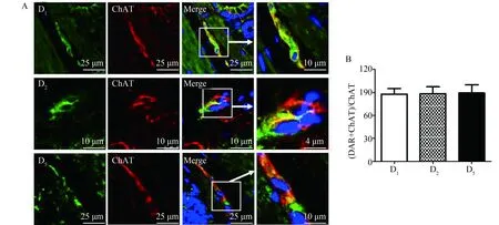
圖2 多巴胺受體在大鼠結腸黏膜下膽堿能神經元上的表達及共存率比較Fig.2 The distribution of dopamine receptors on ChAT-IR neurons in submucosal plexus of rat colon, and comparison of their co-existence
A:green fluorescence staining for dopamine receptors immunoreactivity, red fluorescence staining for ChAT immunoreactivity; B: co-expression rate of D1, D2or D5in ChAT-IR neurons,n=4; ChAT-IR:choline acetyltransferase immunoreactivity; DAR:dopamine receptors.
2.3 多巴胺受體在大鼠結腸的VIP能神經元上的分布
以VIP為VIP能神經元標記,采用免疫熒光雙重染色方法并在激光共聚焦顯微鏡下分別觀察DA受體D1、D2、D5在大鼠遠端結腸黏膜下神經叢內VIP免疫反應陽性神經元上的表達情況。如圖3所示,黏膜下VIP神經元中58.94%±2.245%為D1陽性,52.02%±9.384%為D2陽性,86.21%±2.902%為D5陽性。VIP能神經元上的DA受體D5的表達率顯著高于D1及D2,差異有統計學意義。
A: green fluorescence staining for dopamine receptors immunoreactivity, red fluorescence staining for VIP immunoreactivity; B: co-expression rate of D1, D2or D5in VIP-IR neurons;n=3, *P<0.05;#P<0.05;DAR:dopaminereceptors;VIP-IR:vasoactiveintestinalpeptideimmunoreactivity.
3 討論
研究顯示DA及其受體在結腸肌層、肌間神經叢、黏膜上皮細胞及固有層免疫細胞上均有分布[11],但在大鼠黏膜下神經叢中的分布并不明確。其中肌層及肌間神經叢上的DA受體D1、D5和D2介導DA對結腸動力的抑制作用[12],黏膜層DA受體可在結腸黏膜免疫應答中發揮重要的調節作用[13-14]。本實驗室前期研究[11]顯示DA受體在大鼠結腸黏膜層有D1及D5的表達,本研究顯示大鼠結腸組織的黏膜下層有DA受體D1、D2、D5的表達,且在黏膜下膽堿能神經元及VIP能神經元上均有分布,其中VIP能神經元上D5受體的表達數量顯著高于其他類型。
結腸黏膜下神經叢中分布著大量的神經元,大致分為膽堿能神經元及非膽堿能神經元,在腸道黏液分泌、跨膜離子轉運及免疫調節方面發揮著廣泛的直接或間接的影響作用。其中非膽堿能神經元中,VIP能神經元約占大鼠結腸黏膜下神經元數量的60%[15],VIP與多種神經遞質如NO共存,除參與調節腸道分泌、血流之外,也可通過調節樹突狀細胞功能、抑制巨噬細胞活化以及修復黏膜屏障等作用抑制腸道炎性反應,從而發揮黏膜保護作用[13]。由膽堿能神經元所釋放的ACh同樣具有類似的調節作用,且二者在黏膜分泌功能的調節中還存在協同作用[10,16-17]。
近年來多項研究表明DA及其受體參與炎性腸病的發病機制,但研究結果并不統一:有文獻[18]顯示DA可通過作用于樹突狀細胞上的D5受體促進Th17細胞的分化,促進炎性腸病進程;也有文獻[19]顯示DA可通過作用于D2受體發揮結腸黏膜保護作用。分析可能主要與不同DA受體的細胞分布差異、親和力差異及不同情況下DA濃度改變有關。DA與其受體的親和力各有不同:D3>D5>D4>D2>D1,DA在低濃度時優先結合親和力較高的受體,高濃度時結合較低親和力的受體,進而發揮不同的調節作用[1]。研究[20]顯示炎性腸病時腸道DA濃度顯著降低,因此DA有可能優先與黏膜層免疫細胞表面上的D5受體結合,促進炎性反應進程。而根據本研究結果可推斷,DA還可能與黏膜下神經元上D5受體高親和力結合促進VIP及ACh的釋放,同時發揮抑炎的負反饋調節作用。而D2受體可能在較高濃度時發揮抑制動力及潰瘍生成等作用,具體機制還需深入探討。
綜上,本研究完善了DA受體在大鼠結腸黏膜的形態學分布數據,為深入探討DA對結腸黏膜功能調節機制提供實驗室依據。
[1]PachecoR,ContrerasF,ZoualiM.Thedopaminergicsysteminautoimmunediseases[J].FrontImmunol, 2014, 5:117.
[2] Tolstanova G, Deng X, Ahluwalia A, et al. Role of dopamine and D2 dopamine receptor in the pathogenesis of inflammatory bowel disease[J]. Dig Dis Sci, 2015,60(10):2963-2975.
[3] Zheng L F, Song J, Fan R F, et al. The role of vagal pathway and gastric dopamine in the gastroparesis of rats after 6-hydroxydopamine microinjection in the substantia nigra[J].Acta Physiol (Oxf),2014, 211(2): 434-446.
[4] Feng X Y, Li Y, Li L S, et al. Dopamine D1 receptors mediate dopamine-induced duodenal epithelial ion transport in rats[J].Transl Res,2013,161(6):486-494.
[5] Zhang G H, Zhu J X, Xue H, et al. Dopamine stimulates Cl-absorption coupled with HCO3 -secretion in rat late distal colon[J]. Eur J Pharmacol, 2007,570(1-3):188-195.
[6] Al-Jahmany A A, Schultheiss G, Diener M. Effects of dopamine on ion transport across the rat distal colon[J].Pflugers Arch, 2004,448(6):605-612.
[7] Li Z S, Pham T D, Tamir H, et al. Enteric dopaminergic neurons: definition, developmental lineage, and effects of extrinsic denervation[J]. J Neurosci, 2004, 24(6):1330 -1339.
[8] Li Z S, Schmauss C, Cuenca A, et al. Physiological modulation of intestinal motility by enteric dopaminergic neurons and the D2 receptor: analysis of dopamine receptor expression, location, development, and function in wild-type and lnock-out mice[J]. J Neurosci, 2006,26(10):2798 -2807.
[9] Burns, A J, Hofstra R M. The enteric nervous system: From embryology to therapy[J]. Dev Biol, 2016,417(2):127-128.
[10]Foong J P, Tough I R, Cox H M, et al. Properties of cholinergic and non-cholinergic submucosal neurons along the mouse colon[J]. J Physiol, 2014,592(4):777-793.
[11]Zhang X, Li Y, Liu C, et al. Alteration of enteric monoamines with monoamine receptors and colonic dysmotility in 6-OHDA-induced Parkinson’s disease rats[J].Transl Res,2015, 166(2):152-162.
[12]ZhangX H, Zhang X F, Zhang J Q, et al. β-adrenoceptors, but not dopamine receptors, mediate dopamine-induced ion transport in late distal colon of rats[J].Cell Tissue Res,2008,334(1):25-35.
[13]Chino Y, Fujimura M, Kitahama K, et al. Colocalization of NO and VIP in neurons of the submucous plexus in the rat intestine[J]. Peptides,2002, 23 (12): 2245-2250.
[14]Gonzalez-Rey E, Delgado M. Therapeutic treatment of experimental colitis with regulatory dendritic cells generated with vasoactive intestinal peptide[J]. Gastroenterology, 2006, 131(6): 1799-1811.
[15]Gonzalez-Rey E, Delgado M. Role of vasoactive intestinal peptide in inflammation and autoimmunity[J].Curr Opin Investig Drugs, 2005, 6(11): 1116-1123.
[16]Reddix R, Kuhawara A, Wallace L, et al. Vasoactive intestinal polypeptide: a transmitter in submucous neurons mediating secretion in guinea pig distal colon[J]. J Pharmacol Exp Ther, 1994, 269(3):1124-1129.
[17]Ratcliffe E M, deSa D J, Dixon M F, et al. Intestines choline acetyltransferase (ChAT) immunoreactivity in paraffin sections of normal and diseased intestines[J].J Histochem Cytochem,1998, 46(11): 1223-1231.
[18]Kawano M, Takagi R, Kaneko A, et al. Berberine is a dopamine D1- and D2-like receptor antagonist and ameliorates experimentally induced colitis by suppressing innate and adaptive immune responses[J]. J Neuroimmunol, 2015, 289: 43-55.
[19]Tolstanova G, Deng X, Ahluwalia A, et al. Role of dopamine and D2 dopamine receptor in the pathogenesis of inflammatory bowel disease[J]. Dig Dis Sci, 2015, 60(10): 2963-2975.
[20]Magro F, Vieira-Coelho M A, Fraga S, et al. Impaired synthesis or cellular storage of norepinephrine, dopamine, and 5-hydroxytryptamine in human inflammatory bowel disease[J]. Dig Dis Sci, 2002, 47(1): 216-224.
編輯 孫超淵
Expression and cellular distribution of dopamine receptors in the rat colonic submucosal plexus
Zhang Yue1, 2, Li Yun1*, Zhu Jinxia2, Zhao Wenming1*
(1.DepartmentofImmunology,SchoolofBasicMedicalSciences,CapitalMedicalUniversity,Beijing100069,China; 2.DepartmentofPhysiologyandPathophysiology,SchoolofBasicMedicalSciences,CapitalMedicalUniversity,Beijing100069,China)
Objective Dopamine (DA) plays an important role in the regulation of intestinal motility, secretion and mucosal barrier by binding to its receptors. It has been shown that submucosal neurons are involved in the regulation effect of DA on the mucosa function. However the expressions of DA receptors in submucosal neurons are not clear. The aim of the present study is to investigate the distribution of DA receptors in vasoactive intestinal peptide (VIP) neurons and cholinergic neurons in the submucosa. Methods The mRNA and protein expression of DA receptors was measured by RT-PCR and Western blotting. Immunofluorescent double labeling of DA receptors with submucosal VIP neurons and cholinergic neurons were observed under confocal laser scanning microscope. Results Both the mRNA and protein of DA receptors D1, D2and D5were expressed in the colonic submucosal layer. In the submucosal plexus, D1positive VIP-positive neurons accounting for 58.94%±2.245%, D2for 52.03%±9.384%, and D5for 86.21%±2.902%. The number of D5positive neurons was significantly higher than that of D1and D2. In the submucosal plexus, D1positive cholinergic neurons accounted for 87.75%±7.307%, D2for 88.50%±8.761%, and D5for 89.25%±10.75%, and the distribution of the three types of DA receptors in the cholinergic neurons had no significant difference. Conclusion DA receptors D1, D2and D5are expressed in the submucosal layer of the rat colon. And they all are distributed in the submucosal VIP and cholinergic neurons, and D5expression was higher than D1and D2in VIP-positive neurons. Our study provides a morphological basis for the regulation of DA on the colonic mucosal function.
dopamine; dopamine receptors; submucosal plexus; vasoactive intestinal peptide; acetylcholine
國家自然科學基金(31300954,81370482)。This study was supported by National Natural Science Foundation of China (31300954, 81370482).
時間:2017-06-09 17∶39 網絡出版地址:http://kns.cnki.net/kcms/detail/11.3662.r.20170609.1739.042.html
10.3969/j.issn.1006-7795.2017.03.017]
R392
2016-12-15)
*Corresponding authors, E-mail:liyun666@ccmu.edu.cn; zhao-wenming@163.com