Correlation between age and thickness of Descemet’s membrane in Chinese people
2016-09-02 06:12:21WeiWangLingJuanXuBanLuoXianZhangGuiGangLi
國際眼科雜志 2016年9期
Wei Wang, Ling-Juan Xu, Ban Luo, Xian Zhang, Gui-Gang Li
?
·Original article·
Correlation between age and thickness of Descemet’s membrane in Chinese people
Wei Wang, Ling-Juan Xu, Ban Luo, Xian Zhang, Gui-Gang Li
Foundation items:The National Natural Science Foundation of China (No. 81200661; 81470606); The Nature Science Foundation of Hubei Province (No. 2014CFB973; 2014CFB442)
Department of Ophthalmology, Tongji Hospital, Tongji Medical College, Huazhong University of Science and Technology, Wuhan 430030, Hubei Province, China
Correspondence to:Gui-Gang Li. Department of Ophthalmology, Tongji Hospital, Tongji Medical College, Huazhong University of Science and Technology, Wuhan 430030, Hubei Province, China.guigli@163.com
目的:測量中國人的角膜后彈力層厚度并分析其與年齡的關系。
方法:收集27例角膜標本,年齡范圍從0.2歲到78歲,所有標本經HE染色后行組織切片并通過400倍光學顯微鏡行顯微照相,于每張相片中選取四個測量點并人工標記后彈力層的邊界,通過軟件Motic Images Plus 2.0自動計算其厚度并取平均值。采用線性回歸方法分析角膜后彈力層厚度與年齡的關系。
結果:所有標本的角膜后彈力層厚度為1.78μm到9.30μm,平均4.63±2.00μm。角膜后彈力層厚度與年齡具有高正相關性(r=0.776,P=0.000),線性回歸方程可描述為:角膜后彈力層厚度(μm)=2.010+0.063年齡(歲)。
結論:中國人的角膜后彈力層厚度與年齡之間具有顯著正相關性。
引用:王瑋,徐玲娟,羅班,張憲,李貴剛. 中國人角膜后彈力層厚度與年齡的相關性研究.國際眼科雜志2016;16(9):1599-1602
Abstract
?AIM: To examination the thickness of Chinese Descemet’s membrane and investigate the correlation with age.
?METHODS: Twenty-seven normal corneas with ages ranging from 0.2 to 78y old were collected. All specimens were stained with Hematoxylin and Eosin, and viewed and photographed at ×400 through a calibrated Motic light photomicroscope. Four measurement sites on each specimen micrograph were chosen and the boundary of Descemet’s membrane was designated manually, then the Descemet’s membrane thickness was measured by the software of Motic Images Plus 2.0 automatically. The relationship between Descemet’s membrane thickness and age was analyzed by using linear regression methods.
?RESULTS: The thickness of Chinese Descemet’s membrane various from 1.78 to 9.30 μm, averaged 4.63±2.00 μm. There is a highly significant positive correlation between age and thickness of Descemet’s membrane (r=0.776,P=0.000). The estimated equation that best describes the relationship of thickness with age can be expressed as: Thickness (μm)= 2.010+0.063y.
?CONCLUSION: There is a significant positive association between age and the thickness of Descemet’s membrane in Chinese people.
?cornea; Descemet’s membrane; thickness; age
INTRODUCTION
Descemet’s membrane is the basement membrane of the corneal endothelium[1]. This posterior limiting membrane fulfills important structural and physiologic functions in the cornea[2].
Descemet’s membrane is composed of anterior banded zone and posterior non-banded zone when viewed through scanning electron microscope[3-4]. The anterior banded zone is first secreted in utero by the endothelial cells at about four months’ gestation and acquires recognizable banding by eight months’ gestation. The posterior non-banded zone is secreted by the endothelial cells after birth. Observations in mammals suggest that only the posterior non-banded zone is synthesized continuously throughout adult life[3-6].
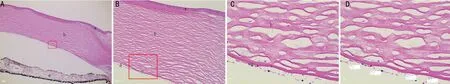
Figure 1Histopathologic appearance of thecornea with hematoxylin and eosin staining(A ×40, B ×100, C ×400, D is same to C but shows the Measurement sites): a is the epithelium cell layer, b is the stroma, c is the Descemet’s membrane, d is the endothelial cell layer, e is the iris.
The normal thickness of human Descemet’s membrane is about 2-3 mm at birth, increasing to approximately 5-6 mm in children and 8-14 mm in adults[3-4]. To our knowledge, there is no morphometric data available that describing the relationship between Descemet’s membrane thickness and age in Chinese people. The purpose of our investigation is to provide a comprehensive Chinese standard derived from measurements of 27 specimens from 0.2 to 78y old.
SUBJECTS AND METHODS
Human corneas were obtained from the Wuhan Red Cross Eye Bank. Informed consent was obtained prior to the study from patients or relatives according to usual procedures, the corneas were managed in accordance with the declaration of Helsinki. Twenty-seven ostensibly normal corneas which ages ranging from 0.2 to 78y old were collected (Table 1). The central parts of 12 corneas were used for transplantation and peripheral parts were used for this study, the other corneas were not suitable for transplantation for HBs positive or cancer disease. Corneas from both male and female patients were included, and only one eye per patient was studied.
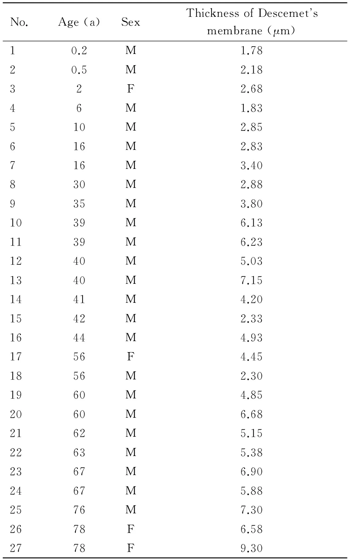
Standard methods were used for fixation and tissue processing of the cornea. Sectioning of the paraffin-embedded tissue was in a meridional plane perpendicular to the corneal surface. Transverse sections (3.0 μm thick) of Descemet’s membrane and adjacent corneal tissue were stained with hematoxylin and eosin(HE), then viewed and photographed at ×400 through a calibrated Motic light photomicroscope(Figure 1). The boundary of Descemet’s membrane was designated manually on the micrographs which allowed for an easy identification of the borders, and the thickness from the anterior boundary adjacent to the stroma to the posterior boundary adjacent to the endothelial cell layer was measured by the software of Motic Images Plus 2.0. This software can measure the thickness automatically by comparing the scale length when the measurement sites were chosen. And the measurement sites choice were not random, as the boundary domelike extension and tangentially sectioned foci were avoided. We took one ×400 micrograph of each cornea specimen, chose four measurement sites on each micrograph, and calculated the average thickness(Figure 2, 3). The relationship between Descemet’s membrane thickness and age was analyzed by using linear regression methods.
Table 1General information for the corneas
No.Age(a)SexThicknessofDescemetsmembrane(μm)10.2M1.7820.5M2.1832F2.6846M1.83510M2.85616M2.83716M3.40830M2.88935M3.801039M6.131139M6.231240M5.031340M7.151441M4.201542M2.331644M4.931756F4.451856M2.301960M4.852060M6.682162M5.152263M5.382367M6.902467M5.882576M7.302678F6.582778F9.30
RESULTS
The thickness of Chinese Descemet’s membrane various from 1.78 to 9.30 μm, averaged 4.63±2.00 μm(n=27). There is a highly significant positive correlation between age and thickness of Descemet’s membrane (r=0.776,P=0.000) (Figure 4). The estimated equation that best describes the relationship of thickness with age can be expressed as: thickness (μm)=2.010+0.063y. But sufficient variation in thickness of Descemet’s membrane between individuals, it’s inaccurate to predict the exact thickness of any people from the age.
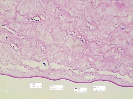
Figure 2The micrograph of 0.2-year-old infant’s Descemet’s membrane with HE staining (×400), the average of 4 measurements of thickness is 1.78 μmThis is the youngest specimen which has the thinnest thickness.
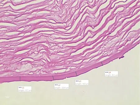
Figure 3The micrograph of 78-year-old woman’s Descemet’s membrane with HE staining (×400), the average of 4 measurements of thickness is 9.30 μmThis is the oldest specimen which has the thickest thickness.
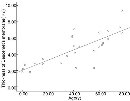
Figure 4Thickness of Descemet’s membrane as a function of age (n=27 specimens, ages from 0.2 to 78y)The estimated equation that best describes the relationship of thickness with age can be expressed as: thickness (μm) =2.010+0.063y.
The specimens which ages ranging from 35 to 44y have an average thickness of 4.98 μm with a standard deviation of 1.54 μm(n=8). And the specimens which ages ranging from 56 to 67y have an average thickness of 5.20 μm with a standard deviation of 1.45 μm(n=8).
DISCUSSION
All we know that the thickness of Descemet’s membrane increases with age[7-8], but this study is the first time to describe the relationship between Descemet’s membrane thickness and age in Chinese people by morphometric data. Descemet’s membrane has a complex structure which consists of collagen components, such as I, III, IV, V, VI, and VIII, and noncollagenous components, such as laminin, heparin sulphate, fibronectin, nidogen, tenascin, andPcomponent, but a complete understanding of the composition of Descemet’s membrane has not yet been achieved[9-11]. Descemet’s membrane is a basement membrane that lies in-between the stroma and the endothelial layer of the cornea, and eosin is a fluorescent red dye which can be used to distinguish Descemet’s membrane from the stroma and the endothelial layer. But the boundary of Descemet’s membrane viewed through light microscope is not clearer than electron microscope, the thickness measurement on light microscope graphs may cause more inaccuracy. And we found if the Descemet’s membrane is thicker, the measurement inaccuracy is bigger. To reduce this inaccuracy, we abandoned some pathologic specimens which tissue is out of normal shape, we chose the measurementsites which the anterior boundary of Descemet’s membrane parallel to the posterior boundary, and we avoided choosing the measurementsites that the anterior or posterior boundary was not clear.
Descemet’s membrane is composed of anterior banded zone and posterior non-banded zone when viewed through scanning electron microscope, and only the posterior non-banded layer thicken with age. But there is no distinction between anterior banded layer and posterior non-banded layer when viewed through light microscope with HE staining. In this paper we analysis the relationship between the entire Descemet’s membrane thickness and age in Chinese people. And the result is similar to previously published, we found a significant positive association between age and the thickness of Descemet’s membrane.
Using transmission electron microscope, Murphyetal[4]measured the average thickness of Descemet’s membrane to be about 10.07±0.99 μm in the ages ranging from 32 to 44y (n=6) and 11.61±2.32 μm in the ages ranging from 55 to 68y (n=10), Johnsonetal[3]measured the average thickness of Descemet’s membrane to be about 10.88±2.49 μm in the ages ranging from 52 to 68y (n=6). But our specimens which ages ranging from 35 to 44y have an average thickness of 4.98±1.54 μm (n=8), and specimens which ages ranging from 56 to 67y have an average thickness of 5.20±1.45 μm (n=8). Compared with the Westerners’ data based on electron microscope, the Chinese data based on light microscope are significantly thinner. There are some elements should be consider which cause this difference in thickness. The Descemet’s membrane boundary lack of precision which we have discussed may cause the variability. Tissue swelling or shrinkage may occur during tissue processing and fixation, and different methods of tissue processing and fixation between electron microscopy and light microscopy may lead to artifacts. Compared Murphy’s data which published based on the average thickness of whole membrane, our study chose only one ×400 micrograph of each cornea specimen to measure the thickness which can’t reflect the average thickness of whole membrane. Because the peripheral part of Descemet’s membrane is thicker than the central part[12-13]. Additionally, the thickness of Descemet’s membrane in Chinese people may be thinner than Westerners, but this point need more specimens to support based on transmission electron microscope.
The most important result of our study is that we have been able to estimate the correlations between the thickness of Descemet’s membrane and age in Chinese people for the first time. Although the thickness of Descemet’s membrane increases with age, the range of thicknesses between individuals is large enough that age can’t be accurately predicted from the thickness in any particular case.
REFERENCES
1 Eghrari AO, Riazuddin SA, Gottsch JD. Overview of the Cornea: Structure, Function, and Development.ProgMolBiolTranslSci2015,134:7-23
2 DelMonte DW, Kim T. Anatomy and physiology of the cornea.JCataractRefractSurg2011;37(3):588-598
3 Johnson DH, Bourne WM,Campbell RJ. The ultrastructure of Descemet’s membrane. I. Changes with age in normal corneas.ArchOphthalmol1982;100(12):1942-1947
4 Murphy C, Alvarado J, Juster R. Prenatal and postnatal growth of the human Descemet’s membrane.InvestOphthalmolVisSci1984;25(12):1402-1415
5 Jun AS, Chakravarti S, Edelhauser HF, Kimos M. Aging changes of mouse corneal endothelium and Descemet’s membrane.ExpEyeRes2006;83(4):890-896
6 Lesueur L, Arne JL, Mignon-Conte M, Malecaze F. Structural and ultrastructural changes in the developmental process of premature infants’ and children’s corneas.Cornea1994;13(4):331-338
7 Peh GS, Beuerman RW, Colman A, Tan DT, Mehta JS. Human corneal endothelial cell expansion for corneal endothelium transplantation: an overview.Transplantation2011;91(8):811-819
8 Chaurasia SS, Champakalakshmi R, Li A, Poh R, Tan XW, Lakshminarayanan R, Lim CT, Tan DT, Mehta JS. Effect of Fibrin Glue on the Biomechanical Properties of Human Descemet’s Membrane.PLoSONE2012;7(5):e37456
9 Levy SG, McCartney AC, Moss J. The distribution of fibronectin andP component in Descemet’s membrane: an immunoelectron microscopic study.CurrEyeRes1995;14(9):865-870
10 Schittny JC, Kresse H, Burri PH. Immunostaining of a heterodimeric dermatan sulphate proteoglycan is correlated with smooth muscles and some basement membranes.HistochemCellBiol1995;103(4):271-279
11 Dua HS, Faraj LA, Said DG, Gray T, Lowe J. Human corneal anatomy redefined: a novel pre-Descemet’s layer (Dua’s layer).Ophthalmology2013;120(9):1778-1785
12 Kafarnik C, Murphy CJ, Dubielzig RR. Canine duplication of Descemet’s membrane.VetPathol2009;46(3):464-473
13 Schl?tzer-Schrehardt U, Bachmann BO, Tourtas T, Torricelli AA, Singh A, González S, Mei H, Deng SX, Wilson SE, Kruse FE. Ultrastructure of the posterior corneal stroma.Ophthalmology2015;122(4):693-699
中國人角膜后彈力層厚度與年齡的相關性研究
王瑋,徐玲娟,羅班,張憲,李貴剛
國家自然科學基金(No.81200661;81470606);湖北省自然科學基金(No.2014CFB973;2014CFB442)
430030中國湖北省武漢市華中科技大學同濟醫學院附屬同濟醫院眼科)
王瑋,畢業于華中科技大學,7年制碩士,主治醫師,研究方向:眼表疾病。
李貴剛,畢業于華中科技大學,博士,副主任醫師,副教授,研究方向:眼表疾病. guigli@163.com
角膜;后彈力層;厚度;年齡
10.3980/j.issn.1672-5123.2016.9.01
Wang W, Xu LJ, Luo B, Zhang X, Li GG. Correlation between age and thickness of Descemet’s membrane in Chinese people.GuojiYankeZazhi(IntEyeSci) 2016;16(9):1599-1602
2015-12-27Accepted: 2016-06-23
