天胡荽的解剖和屏障結構特征研究
2016-01-27 07:06:37楊朝東李守峰姚蘭艾訓儒蔡小東張霞
草業學報 2015年7期
楊朝東,李守峰,姚蘭,艾訓儒,蔡小東,張霞*
(1.長江大學園藝園林學院, 湖北 荊州 434025;2.湖北省保康縣林業局, 湖北 保康441600;
3.湖北民族學院林學園藝學院,湖北 恩施445000)
天胡荽的解剖和屏障結構特征研究
楊朝東1,李守峰2,姚蘭3,艾訓儒3,蔡小東1,張霞1*
(1.長江大學園藝園林學院, 湖北 荊州 434025;2.湖北省保康縣林業局, 湖北 保康441600;
3.湖北民族學院林學園藝學院,湖北 恩施445000)
摘要:利用光學顯微鏡和熒光顯微鏡對天胡荽進行了解剖學和組織化學研究,結果表明,1)天胡荽不定根為初生結構,具二原型維管柱、內皮層、皮層、外皮層和表皮。2)莖、花柄和葉主要為初生結構,除了莖和花柄維管束具次生結構,具表皮、厚角組織、皮層、內皮層、維管束和髓;莖具誘導型通氣組織。3)天胡荽不定根的屏障結構包括內側的有凱氏帶且栓質化的內皮層,外側的有凱氏帶且栓質化的外皮層和緊鄰外側具擴散狀栓質層的表皮細胞;匍匐莖、花柄和葉柄具相似的質外體屏障結構,一是內側的有凱氏帶且栓質化的內皮層,二是外側的表皮外角質層,但花柄和葉柄有凱氏帶的細胞,并不栓質化;葉片的僅為表皮外角質層。4)天胡荽的解剖和屏障結構特征是其適應多種水濕環境的結構基礎。該研究為今后選擇濕地生態修復植物提供線索。
關鍵詞:天胡荽;解剖結構;質外體屏障結構;組織化學;初生結構
DOI:10.11686/cyxb2014246
Yang C D, Li S F, Yao L, Ai X R, Cai X D, Zhang X. A study of anatomical structure and apoplastic barrier characteristics ofHydrocotylesibthorpioides. Acta Prataculturae Sinica, 2015, 24(7): 139-145.
楊朝東, 李守峰, 姚蘭, 艾訓儒, 蔡小東, 張霞. 天胡荽的解剖和屏障結構特征研究. 草業學報, 2015, 24(7): 139-145.
http://cyxb.lzu.edu.cn
收稿日期:2014-05-16;改回日期:2014-06-10
基金項目:湖北省教育廳項目(Q2014310)資助。
作者簡介:楊朝東(1971-),男,湖北巴東人,副教授,博士。E-mail:chaodongyang@aliyun.com
通訊作者*Corresponding author. E-mail:zhang.yang07@aliyun.com
Abstract:The anatomy and histochemistry of Hydrocotyle sibthorpioides were investigated under light and fluorescence microscopy in this research. The findings were: 1) The adventitious root of H. sibthorpioides consisted of proto-xylem and proto-phloem, superceded by diarch primary vasculature of metaxymlem and metaphloem, surrounded by endodermis, then cortex, exodermis and epidermis. 2) Stolon, pedicel and leaf were primary structures with epidermis, collenchyma, cortex, endodermis, vascular bundle and pith, except that vasculature of the stolon and pedicel had secondary structures, and stolon had induced aerenchyma. 3) The apoplastic barriers in adventitious roots of H. sibthorpioides included the inner layers of endodermis with Casparian bands and suberin, the outer layers of exodermis with Casparian bands and suberin and the epidermis with suberin. The stolons, pedicels and petioles had similar apoplastic barriers, including the inner layer of endodermis with Casparian bands and suberin, and the outer layer of the cuticle, but the endodermis had no suberin in pedicels and petioles; leaf blades had only a cuticular apoplastic barrier. 4) The anatomical structure and apoplastic barrier characters are important structural determinants of adaptation of H. sibthorpioides to variably-moist environments. The implications of this research for selection of plant species to restore degraded wetlands are discussed.
A study of anatomical structure and apoplastic barrier characteristics ofHydrocotylesibthorpioides
YANG Chao-Dong1, LI Shou-Feng2, YAO Lan3, AI Xun-Ru3, CAI Xiao-Dong1, ZHANG Xia1*
1.TheCollegeofGardeningandHorticulture,YangtzeUniversity,Jingzhou434025,China; 2.BaokangCountryForestryBureau,Baokang441600,China; 3.TheCollegeofForestryandHorticulture,HubeiMinzuUniversity,Enshi445000,China
Key words:Hydrocotylesibthorpioides; anatomy; apoplastic barriers; histochemistry; primary structure
天胡荽(Hydrocotylesibthorpioides)為傘形科天胡荽屬植物,廣泛分布于我國南方。天胡荽為較細小的多年生雙子葉植物,能在干旱、濕地和完全水淹條件下正常生活,對水濕具很強的適應能力,既作為新型草坪,也有很高的藥用價值[1-2],但其適應各種水濕條件的解剖結構和質外體屏障結構尚無研究報道。
濕地植物適應濕地缺氧環境必須具備兩類重要結構,即通氣組織和質外體屏障結構[3-7]。屏障結構是由植物體各器官內、外皮層初生壁的凱氏帶,或次生壁栓質化和木質化,以及植物體表角質層組成的保護組織[3,8-16]。質外體屏障和細胞膜水孔蛋白是控制植物內部以及與環境的水、離子和氧氣擴散和交換的重要結構[4,8,10-14]。濕地植物體內氣腔主要包括根中通氣組織,莖中髓腔和皮層通氣組織,保障植物淹沒脅迫時儲藏和輸導氧氣[16-18]。
經典植物形態解剖學主要在光學顯微鏡和電鏡下,觀察植物細胞組織結構和胞內細胞器超微構造,也有染色,其目的是便于觀察胞壁和細胞器的形態和邊界[19-21]。近些年,隨著組織化學、熒光顯微鏡的應用、植物細胞發育及其分子機理的研究,植物解剖學取得了以下幾方面的重要進展,植物的皮層、內皮層、外皮層和皮下層的概念與經典植物解剖學所指的結構和意義完全不同。一是根尖細胞發育生物學及其基因表達等的研究結果表明,擬南芥(Arabidopsisthaliana)根尖內皮層和皮層具有共同的原始細胞(CEI),表皮具獨立來源的原始細胞;水稻(Oryzasativa)根尖皮層、內皮層、外皮層和表皮具共同的原始細胞(CEEI)[22]。二是擬南芥根內皮層凱氏帶形成過程中,有凱氏帶膜蛋白家族(CASPs)參與,同時說明凱氏帶中具有蛋白質,該蛋白與凱氏帶在初生壁上也構成類似于動物上皮組織細胞間僅蛋白質參與的緊密連接,具有保護作用[23-24]。三是擬南芥根內皮層凱氏帶應具備凱氏帶結構、圍繞維管束的拓撲學結構和SCR基因的表達三大要素[25]。四是凱氏帶部位先有凱氏帶膜蛋白家族出現,隨后有木質素沉積,再有木栓質沉積[26]。五是依據根內皮層細胞初生壁和次生壁出現凱氏帶之前、凱氏帶、栓質化和木質化的組織化學出現先后順序,把內皮層的發育分為前內皮層階段、第Ⅰ、Ⅱ 和Ⅲ階段,或者停留在某發育階段[8]。因此,認為植物內皮層或外皮層一定具有凱氏帶,凱氏帶不僅有木栓質和木質素,還含有蛋白質。植物的內皮層、皮層、外皮層和表皮是同等意義結構。皮下層是指表皮下有細胞形態分化的結構,或含有外皮層或和厚壁層分化的結構。皮層是內皮層和外皮層之間的薄壁細胞組織。
三峽庫區消落帶的生態修復問題,雖然從濕地植物的形態結構與生理、種群分布格局等方面已取得進展,但解決該生態修復問題還有諸多工作需要開展。本研究試圖從天胡荽的解剖結構、組織化學特征和質外體屏障結構,為今后三峽庫區消落帶的生態修復尋找適合耐水淹植物提供線索。
1材料與方法
天胡荽實驗材料準備,于2013年3月底采集湖北荊州長江大學西校區附近野生植株,用農田土壤栽培于花盆中,置于旱地花盆口部與地面平齊,在自然的光和水分條件下生長,一部分花盆作為旱生處理;另一部分花盆在6月中旬,模擬自然洪水脅迫,將有完整天胡荽植株的花盆沉入自來水中,8月中旬取出,水中植株未見開花。取旱生和水淹下樣本進行解剖學實驗。取完整不定根約長75 mm左右,完整匍匐莖約長350 mm左右,具4~5個節。不定根用FAA固定,匍匐莖、花柄和葉采用新鮮材料。
在立體解剖鏡(JNOEC JSZ6, China)下,用雙面刀片切片距根尖10, 30, 50, 70 mm;莖第1節間和第4節間,花柄中部,葉柄中部和葉片基部主脈。硫氫酸黃連素-苯氨蘭對染切片確定細胞壁木質化和凱氏帶,凱氏帶呈現生動黃色(vivid yellow), 木質化細胞壁呈現呆滯黃色(stagnant yellow),如木質部[12,27],蘇丹紅7B染色切片檢測栓質化細胞壁, 呈現紅色,具有很強的組織特異性,而蘇丹Ⅲ或Ⅳ常將細胞內的脂肪也染成紅色,特異性差,造成栓質化胞壁不易識別[28]。蘇丹紅7B染色切片在萊卡光學顯微鏡(Leica DME)下觀察,用數碼相機(Nikon E5400, Japan)拍照記錄圖片。硫氫酸黃連素-苯氨蘭對染切片在熒光顯微鏡(Olympus IX71)藍色激發光下觀察,用數碼相機(RZ200C-21, China)拍照記錄圖片,有關其他操作可參考Yang等[16]。圖片主要用 Photoshop 7.0軟件處理,加標尺,調整圖片亮度和對比度,以及合成圖片等。
2結果與分析
2.1 不定根的解剖結構和組織化學
旱生和水淹下天胡荽的不定根有相似的解剖結構和組織化學發育過程。距天胡荽根尖10和30 mm, 內、外皮層開始形成微弱的凱氏帶,部分細胞壁栓質化(圖1A,B),皮層具1~2層細胞,無通氣組織,表皮細胞壁無擴散狀栓質層,二原型初生木質部內有后生木質部發育,其兩側為韌皮部。距根尖50 mm, 除內皮層的通道細胞外,內、外皮層具強烈的凱氏帶,并且細胞壁栓質化;部分表皮細胞壁開始出現擴散狀栓質層[29](圖1C,D)。在根基部(距根尖70 mm),內、外皮層細胞具凱氏帶,并強烈栓質化,皮層僅具2層細胞,無通氣組織,維管柱停留在初生結構階段(圖1E,F);內皮層、皮層、外皮層和表皮細胞不脫落;表皮細胞壁均具擴散狀栓質層。在不定根的各發育階段,已分化的不定根皮層未脫落,維管柱處于初生結構狀態。不定根中質外體屏障結構包括兩部分,一是有凱氏帶且栓質化的內皮層,二是有凱氏帶且栓質化的外皮層和緊鄰外側具擴散狀栓質層的表皮細胞。
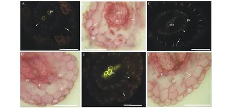
圖1 天胡荽不定根的顯微結構Fig.1 Photomicrographs of H. sibthorpioides adventitious roots 標尺=50 μm. A~B. 距根尖10 mm橫切, A. BAB, 內皮層凱氏帶(箭頭), 外皮層凱氏帶(箭), B. SR7B, 內皮層栓質化(箭頭), 外皮層栓質化(箭); C~D. 距根尖50 mm橫切, C. BAB, 內皮層凱氏帶(箭頭), 外皮層凱氏帶(箭), D. SR7B, 內皮層栓質化(箭頭), 外皮層栓質化(箭); E~F. 距根尖70 mm橫切, E. BAB, 內皮層凱氏帶(箭頭), 外皮層凱氏帶(箭), F. SR7B, 內皮層栓質化(箭頭), 外皮層栓質化(箭)。 BAB. 硫氫酸黃連素-苯氨蘭對染; co. 皮層; ep. 表皮; mx. 后生木質部; p. 韌皮部; pc. 通道細胞; px. 原生木質部; SR7B. 蘇丹紅7B染色; sx. 次生木質部; vb. 維管束。Scale bars=50 μm. A-B. Transverse sectioned at 10 mm from apex, A. BAB, endodermis (arrowheads), exodermis (arrows), B. SR7B, endodermis (arrowheads), exodermis (arrows); C-D. Sectioned at 50 mm, C. BAB, endodermis (arrowheads), exodermis (arrows), D. SR7B, endodermis (arrowheads), exodermis (arrows); E-F. Sectioned at 70 mm, E. BAB, endodermis (arrowheads), exodermis (arrows), F. SR7B, endodermis (arrowheads), exodermis (arrows). BAB. berberine hemisulfate-aniline blue stained, co. cortex; ep. epidermis; mx. metaxylem; p. phloem; pc. passage cell; px. protoxylem; SR7B. Sudan red 7B stained; sx. secondary xylem; vb. vascular bundle.
2.2 匍匐莖、花柄和葉的解剖結構和組織化學
旱生天胡荽匍匐幼莖的解剖結構自外向內依次由表皮、皮層、維管束、髓射線和髓構成。束狀初生維管束相互獨立,之間為薄壁細胞,呈輪狀排列;維管束可見初生木質部導管。皮層細胞暫無分化,表皮下為1~2層細胞的厚角組織,表皮外有角質層(圖2A,B)。
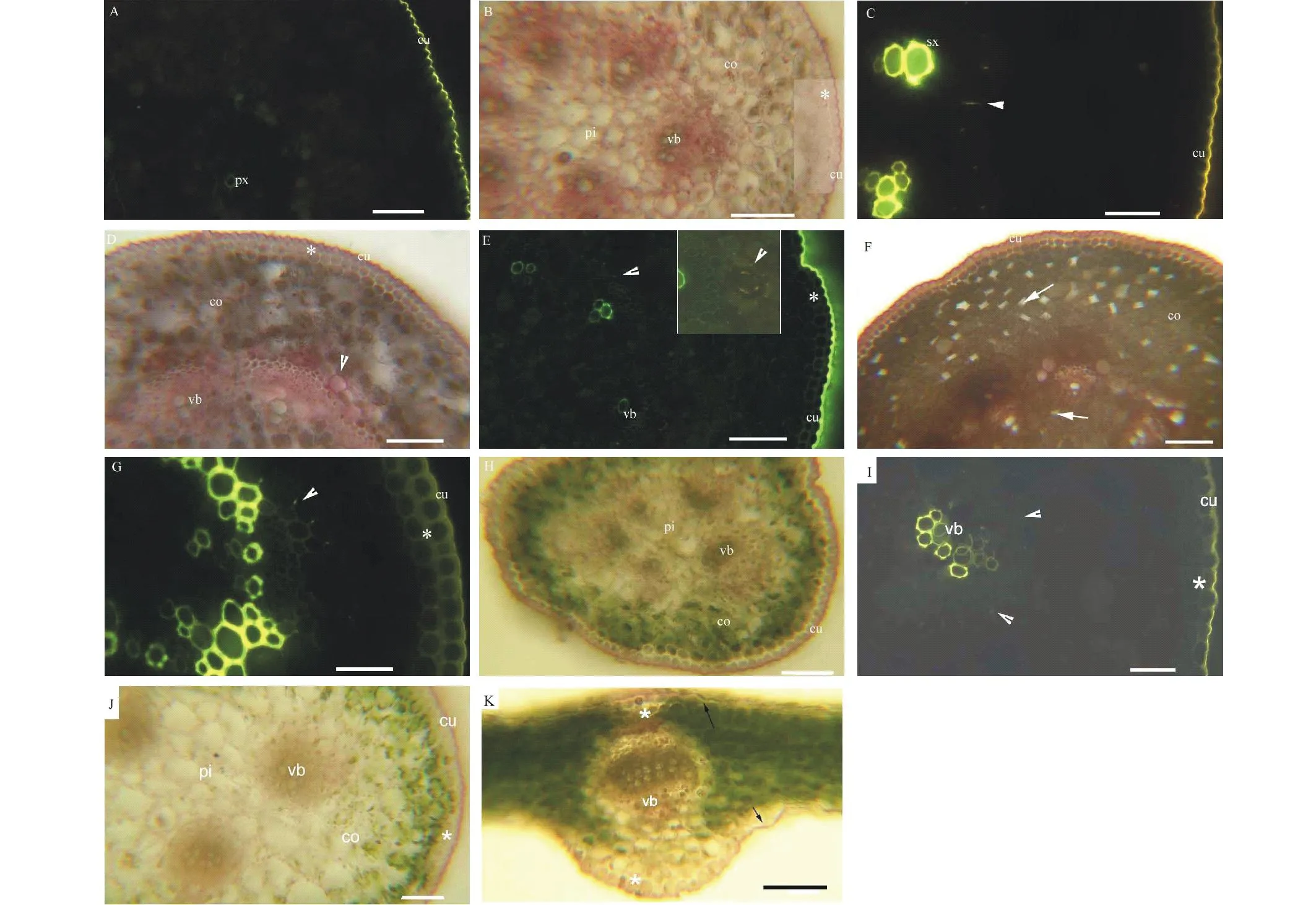
圖2 天胡荽匍匐莖、花柄和葉柄的顯微結構Fig.2 Photomicrographs of H. sibthorpioides stolon, pedicel, petiole and leaf blade 標尺=100 μm. A~B. 幼莖橫切, 厚角組織(星號), A. BAB, B. SR7B; C~D. 老莖橫切, C. BAB, 內皮層凱氏帶(箭頭), D. SR7B, 內皮層栓質化(箭頭), 厚角組織(星號); E~F. 水下老莖橫切, E. BAB, 內皮層凱氏帶(箭頭), 厚角組織(星號), 插入圖為內皮層(箭頭), F. SR7B, 內皮層栓質化(箭頭), 擴大型通氣組織(箭), 厚角組織(星號); G~H. 花柄橫切, G. BAB, 內皮層凱氏帶(箭頭), 厚角組織(星號), H. SR7B, 厚角組織(星號); I~J. 葉柄橫切, I. BAB, 內皮層凱氏帶(箭頭), J. SR7B, 厚角組織(星號). K. 葉片主脈橫切, SR7B, 厚角組織(星號), 角質層(箭)。BAB. 硫氫酸黃連素-苯氨蘭對染; co. 皮層; cu. 角質層; vb. 維管束; pi. 髓部; px. 原生木質部; SR7B. 蘇丹紅7B染色; sx. 次生木質部。Scale bars=100 μm. A-B. Transverse sectioned at the first internode of young stolon, collenchyma (asterisk), A. BAB, B. SR7B; C-D. Transverse sectioned at the 4th internode of aged stolon, C. BAB, endodermis (arrowheads), D. SR7B, endodermis (arrowheads), collenchyma (asterisk); E-F. Transverse sectioned at the 4th internode of aged stolon submerged, E. BAB, endodermis (arrowheads), collenchyma (asterisk), inset is endodermis (arrowheads), F. SR7B, endodermis (arrowheads), expansigeny aerenchyma (arrows), collenchyma (asterisk); G-H. Transverse sectioned the middle of pedicel, G. BAB, endodermis (arrowheads), collenchyma (asterisk), H. SR7B, collenchyma (asterisk); I-J. Transverse sectioned the middle of petiole, I. BAB, endodermis (arrowheads), J. SR7B, collenchyma (asterisk); K. Transverse sectioned the base main vein of leaf blade, SR7B, collenchyma (asterisk), cuticle (arrows). BAB. berberine hemisulfate-aniline blue stained, co. cortex; cu. cuticle; vb. vascular bundle; pi. pith; px. protoxylem; SR7B. Sudan red 7B stained; sx. secondary xylem.
老莖中,表皮外角質層增厚;表皮下厚角組織細胞壁進一步加厚,細胞層數增加;皮層內側有具凱氏帶的內皮層分化,內皮層且栓質化;維管束有次生木質部出現(圖2C,D)。水淹下老莖與旱生的比較,在皮層和髓部出現擴大型通氣組織(圖2E,F),而旱生老莖內無通氣組織。花柄的解剖結構和組織化學與老莖的類似,但內皮層未栓質化;厚角組織下數層細胞含葉綠體(圖2G,H)。
旱生和水淹下天胡荽的葉解剖結構和組織化學相似,葉柄由外而內及其特點依次為:表皮外有角質層,表皮下為厚角組織;厚角組織下數層細胞含葉綠體,皮層,皮層內方為3個相互獨立的維管束,內部為髓部(圖2I,J);每個維管束鞘細胞具凱氏帶,但未栓質化。葉片主脈中間為維管束,維管束上下兩側為厚角組織,最外為角質層,主脈兩側葉片為葉肉細胞(圖2K)。
天胡荽匍匐莖、花柄和葉具相似的質外體屏障結構,一是有凱氏帶且栓質化的內皮層,二是表皮外角質層,但花柄和葉柄有凱氏帶的細胞,并不栓質化;葉片的質外體屏障僅為表皮外角質層。
3討論
3.1 天胡荽的解剖結構
在天胡荽的各發育階段,不定根、匍匐莖、花柄和葉的結構以初生結構為主,除了匍匐莖和花柄維管束具次生結構。不定根的結構為二原型維管柱,具有表皮,皮層內側細胞分化為內皮層,外側為外皮層。莖和花柄的結構為表皮、皮層,皮層外側為厚角組織、皮層內側分化為內皮層,初、次生維管束環狀排列,內部有髓。花柄厚角組織下數層細胞具葉綠體。葉柄的維管束散生,厚角組織下數層細胞具葉綠體。葉片主脈中間為維管束,上下兩側為厚角組織,葉片具葉肉細胞。天胡荽與海邊生長的藥用同屬植物H.umbellate[30],在莖、花柄和葉片的解剖結構相似。
常見雙子葉植物老根的維管柱后期有次生結構發育,皮層脫落,由維管柱鞘細胞發育而來的木栓層代替皮層的保護功能。而天胡荽不定根的皮層僅2層細胞,皮層不脫落,無通氣組織;也不同于常見濕地植物的根皮層具發達通氣組織,如水稻、蘆葦(Phragmitesaustralis)等[4,7,11-14,16-18]。
常見雙子葉植物莖的次生木栓層代替皮層的保護功能,天胡荽莖維管束具次生結構,皮層內側有內皮層分化,表皮下有厚角組織。天胡荽莖在水淹脅迫條件下出現通氣組織,而濕地植物莖中通氣組織為組成型[7,9,16-17,30]。
3.2 不定根屏障結構
天胡荽不定根的質外體屏障由兩部分組成,內側為具凱氏帶且栓質化的內皮層,外側為具凱氏帶且栓質化的外皮層和具擴散狀栓質層的表皮細胞。植物根中內皮層都為單層細胞,而外皮層隨物種不同有單層、2層和多層細胞之分[6,12]。天胡荽、紅樹林和亞馬遜雨林耐淹植物幼根等的外皮層為單層細胞[31-32],而水稻、玉米(Zeamays)、蘆葦、狗牙根(Cynodondactylon)等的外皮層為2層和多層細胞[11-14,16,18]。此外天胡荽與雙穗雀稗(Paspalumdistichum)、玉米、菰(Zizanialatifolia)等的不定根表皮具擴散狀栓質層[18,29,33-34]。盡管天胡荽不定根的外側屏障結構為單層細胞外皮層和具栓質層的表皮細胞,對天胡荽適應各種水濕環境具有重要的保護作用。
3.3 莖、花柄和葉屏障結構
天胡荽匍匐莖和花柄具相似的質外體屏障結構為內側的內皮層和表皮的角質層,葉柄的維管束鞘細胞具凱氏帶,花柄和葉柄具凱氏帶的細胞不栓質化,葉片的僅為角質層。雙穗雀稗、牛鞭草(Hemarthriaaltissima)、狗牙根和菰莖的屏障結構為外側的角質層和表皮下栓質化和木質化的周緣厚壁層,或者有內側栓質化和木質化的厚壁組織,雙穗雀稗表皮下具凱氏帶[7,16,18]。天竺葵莖的屏障結構為具凱氏帶的木栓層[10],香蒲屬植物莖具內皮層[9],水生毛茛的莖具內皮層,葉柄散生維管束鞘細胞具凱氏帶[15],水稻中胚軸為散生維管束鞘細胞具凱氏帶、第一節莖的內皮層和其厚壁組織內散生維管束鞘細胞具凱氏帶[35]。天胡荽匍匐莖、花柄和葉的屏障結構是其忍耐多種水濕環境的重要特征。
3.4 天胡荽解剖結構和屏障結構的生態學意義
天胡荽解剖結構主要為初生結構,除了匍匐莖和花柄維管束具次生結構,水淹誘導莖中形成通氣組織。屏障結構包括不定根的內、外皮層和表皮栓質層;莖和花柄的內皮層、葉柄具凱氏帶的維管束鞘細胞和角質層。天胡荽的解剖結構和屏障結構特征是其適應多種水濕環境的結構基礎,是篩選適合用于三峽庫區消落帶生態修復植物的重要依據。
References:
[1]Gu Z H, Xiang G H, Peng Y L. An introduction test ofHydrocotylesibthorpoioides, a new type of lawn grass. Guizhou Agricultural Sciences, 2009, 37(10): 19-20.
[2]Zhang L, Zhang D Z. Research advance on progress ofHydrocotylesibthorpoioides. Journal of Modern Food and Pharmaceuticals, 2007, 17(1): 15-17.
[3]Armstrong J, Jones R E, Armstrong W. Rhizome phyllosphere oxygenation in Phragmites and other species in relation to redox potential, convective gas flow, submergence and aeration pathways. New Phytologist, 2006, 172: 719-731.
[4]Colmer T D, Gibberd M R, Wiengweera A,etal. The barrier to radial oxygen loss from roots of rice (OryzasativaL.) is induced by growth in stagnant solutions. Journal of Experimental Botany, 1998, 49: 1431-1436.
[5]Greenway H, Armstrong W, Colmer T D. Conditions leading to high CO2(>5 kPa) in waterlogged-flooded soils and possible effects on root growth and metabolism. Annals of Botany, 2006, 98: 9-32.
[6]Yang C D, Zhang X, Liu G F,etal. Progress on the structure and physiological functions of apoplastic barriers in root. Bulletin of Botanical Research, 2013, 33(1):114-119.
[7]Yang C D, Zhang X. Permeability and supplement structures of stems ofPaspalumdistichum. Bulletin of Botanical Research, 2013, 33(5): 564-568.
[8]Enstone D E, Peterson C A, Ma F. Root endodermis and exodermis: structure, function, and responses to the environment. Journal of Plant Growth Regulation, 2003, 21: 335-351.
[9]Mc Manus H A, Seago Jr J L, Marsh L C. Epifluorescent and histochemical aspects of shoot anatomy ofTyphalatifoliaL.,TyphaangustifoliaL. andTyphaglaucaGodr. Annals of Botany, 2002, 90: 489-493.
[10]Meyer C J, Peterson C A. Casparian bands occur in the periderm ofPelargoniumhortorumstem and root. Annals of Botany, 2011, 107: 591-598.
[11]Ranathunge K, Lin J, Steudle E,etal. Stagnant deoxygenated growth enhances root suberization and lignifications, but differentially affects water and NaCl permeabilities in rice (OryzasativaL.) roots. Plant Cell Environment, 2011, 34: 1223-1240.
[12]Seago Jr. J L, Peterson C A, Enstone D E,etal. Development of the endodermis and hypodermis ofTyphaglaucaGodr. andT.angustifoliaL. roots. Canadian Journal of Botany, 1999, 77: 122-134.
[14]Soukup A, Armstrong W, Schreiber L,etal. Apoplastic barriers to radial oxygen loss and solute penetration: a chemical and functional comparison of the exodermis of two wetland species,PhragmitesaustralisandGlyceriamaxima. New Phytologist, 2007, 173: 264-278.
[15]Vecchia F D, Cuccato F, Rocca N L,etal. Endodermis-like sheaths in the submerged freshwater macrophyteRanunculustrichophyllusChaix. Annals of Botany,1999, 83: 93-97.
[16]Yang C D, Zhang X, Zhou C Y,etal. Root and stem anatomy and histochemistry of four grasses from the Jianghan Floodplain along the Yangtze River, China. Flora, 2011, 206: 653-661.
[17]Seago Jr. J L, Marsh L C, Stevens K J,etal. A re-examination of the root cortex in wetland flowering plants with respect to aerenchyma. Annals of Botany, 2005, 96: 565-579.
[18]Yang C, Zhang X, Li J,etal. Anatomy and histochemistry of roots and shoots in wild rice (ZizanialatifoliaGriseb.). Journal of Botany, 2014, 2014: http://dx.doi.org/10.1155/2014/181727.
[19]Liu L L, Yang X, Gao T P,etal. A study on the attractive function of different floral structures inTrolliusranunculoides(Ranunculaceae). Acta Prataculturae Sinica, 2013, 22(3): 190-195.
[20]Lu Q L, Chai S X, Zhang L J,etal. Contribution of winter wheat leaf and non-leaf organs to grain weight. Acta Prataculturae Sinica, 2013, 22(5): 165-174.
[21]Zhang X X, Liu M, Cheng X Y,etal. Comparative study of the morphological and anatomical features ofLinderniaprocumbensin different ecological environments (Lindernuacea). Acta Prataculturae Sinica, 2014, 23(2): 235-242.
[22]Pauluzzi G, Divol F, Puig J,etal. Surfing along the root ground tissue gene network. Developmental Biology, 2012, 365: 14-22.
[23]Roppolo D, De Rybel B, Tendon V D,etal. A novel protein family mediates Casparian strip formation in the endodermis. Nature, 2011, 473: 380-383.
[24]Alassimone J, Roppolo D, Geldner N,etal. The endodermis-development and differentiation of the plant’s inner skin. Protoplasma, 2012, 249(3): 433-443.
[25]Geldner N. The endodermis. Annual Review in Plant Biology, 2013, 64: 531-558.
[26]Naseer S, Leea Y, Lapierre C,etal. Casparian strip diffusion barrier inArabidopsisis made of a lignin polymer without suberin. Proceedings of the National Academy of Science USA, 2012, 109: 10101-10106.
[27]Brundrett M C, Enstone D E, Peterson C A. A berberine-aniline blue fluorescent staining procedure for suberin, lignin and callose in plant tissue. Protoplasma, 1988, 146: 133-142.
[28]Brundrett M C, Kendrick B, Peterson C A. Efficient lipid staining in plant material with Sudan red 7B or Fluorol yellow 088 in polyethylene glycol-glycerol. Biotechnic and Histochemistry, 1991, 66: 111-116.
[29]Schreiber L, Franke R B. Endodermis and Exodermis in Roots eLSM. Chichester: John Wiley and Sons Ltd. 2011. doi: 10.1002/9780470015902. a0002086. pub2.
[30]Martins M B G, Marconi A P, Cavalheiro A J,etal. Anatomical and chemical characterization of the leaf and root system ofHydrocotyleumbellata(Apiaceae). Brazilian Journal of Pharmacognosy, 2008, 18(3): 402-414.
[31]De Simone O, Haase K, Müller E,etal. Apoplastic barriers and oxygen transport properties of hypodermal cell walls in roots from four Amazonian tree species. Plant Physiologist, 2003, 132: 206-217.
[32]Krishnamurthy P, Jyothi-Prakash P A, Qin L,etal. Role of root hydrophobic barriers in salt exclusion of a mangrove plantAvicenniaofficinalis. Plant, Cell Environment, 2014, doi: 10.1111/pce.12272.
[33]Abiko T, Kotula L, Shiono K,etal. Enhanced formation of aerenchyma and induction of a barrier to radial oxygen loss in adventitious roots ofZeanicaraguensiscontribute to its waterlogging tolerance as compared with maize (Zeamaysssp. mays). Plant Cell Environment, 2012, 35: 1618-1630.
[34]Zhang X, Yang C D, Ning G G. The developmental comparison of apoplastic barriers inCynodondactylonandPaspalumdistichumroots. Hubei Agricultural Sciences, 2013, 52(20): 4991-4994.
[35]Watanabe H, Saigusa M, Morita S. Identification of Casparian bands in the mesocotyl and lower internodes of rice (OryzasativaL.) seedlings using fluorescence microscopy. Plant Production Science, 2006, 9: 390-394.
參考文獻:
[1]顧振華, 向國紅, 彭友林. 天胡荽新型草坪引種試驗研究. 貴州農業科學, 2009, 37(10): 19-20.
[2]張蘭, 張德志. 天胡荽的研究進展. 現代食品與藥品雜志, 2007, 17(1): 15-17.
[6]楊朝東, 張霞, 劉國鋒, 等. 植物根中質外體屏障結構和生理功能研究進展. 植物研究, 2013, 33(1): 114-119.
[7]楊朝東, 張霞. 雙穗雀稗(Paspalumdistichum)通透性生理和莖解剖結構補充研究. 植物研究, 2013, 33(5): 564-568.
[19]劉樂樂, 楊曉, 高天鵬, 等. 毛茛狀金蓮花花部結構的吸引功能. 草業學報, 2013, 22(3): 190-195.
[20]魯清林, 柴守璽, 張禮軍, 等. 冬小麥葉片和非葉器官對粒重的貢獻. 草業學報, 2013, 22(5): 165-174.
[21]張欣欣, 劉玫, 程薪宇, 等. 不同生境下陌上菜的形態解剖學比較. 草業學報, 2014, 23(2): 235-242.
[34]張霞, 楊朝東, 寧國貴. 狗牙根和雙穗雀稗根中質外體屏障結構發育過程的比較研究. 湖北農業科學, 2013, 52(20): 4991-4994.