獼猴神經干細胞的誘導分化、干性維持及同種腦內移植
2012-12-25 06:40:48董錦潤郭立云瞿家桂戚仁莉王文超肖春杰王正波
Zoological Research 2012年1期
董錦潤, 郭立云, 瞿家桂, 戚仁莉, 王文超, 肖春杰, 王正波,*
(1. 中國科學院生物物理研究所 腦與認知國家重點實驗室, 北京 100101; 2. 中國科學院昆明動物研究所, 云南 昆明 650223; 3. 云南大學 生命科學院, 云南 昆明 650223; 4. 昆明醫學院第四附屬醫院 眼科, 云南 昆明 650021; 5. 中國科學技術大學 生命科學學院, 安徽 合肥230026)
獼猴神經干細胞的誘導分化、干性維持及同種腦內移植
董錦潤1,2,3,+, 郭立云4,+, 瞿家桂1,2,5, 戚仁莉1,2,5, 王文超1,2, 肖春杰3,*, 王正波1,2,*
(1. 中國科學院生物物理研究所 腦與認知國家重點實驗室, 北京 100101; 2. 中國科學院昆明動物研究所, 云南 昆明 650223; 3. 云南大學 生命科學院, 云南 昆明 650223; 4. 昆明醫學院第四附屬醫院 眼科, 云南 昆明 650021; 5. 中國科學技術大學 生命科學學院, 安徽 合肥230026)
為探索獼猴神經干細胞分化及特性維持, 推進神經干細胞臨床應用研究, 該實驗以綠色熒光蛋白(green fluorescence protein, GFP)為標記探討獼猴胚胎干細胞向玫瑰花環(rosettes)結構神經干細胞的分化及其堿性成纖維細胞生長因子(basic fibroblast growth factor, bFGF)和表皮生長因子(epidermal growth factor, EGF)的擴增培養。結果表明:1)建立了穩定高效的獼猴神經干細胞分化體系, 在該分化體系下, GFP標記獼猴胚胎干細胞在分化的第12天時, 95%以上的細胞分化為神經干細胞; 2)分化得到的Rosettes結構神經干細胞經bFGF/EGF擴增后, 能夠較好地維持其Rosettes結構; 3)經bFGF/EGF擴增后的rosettes結構神經干細胞移植到獼猴腦內后能夠較好的存活并向神經元分化,即bFGF/EGF擴增培養能較好地維持Rosettes結構的神經干細胞, 且移植到獼猴腦內的該細胞亦能夠較好地存活并向神經元分化,該結果為神經干細胞應用于臨床提供了基礎理論依據。
獼猴;神經干細胞;玫瑰花環結構;堿性成纖維細胞生長因子;表皮生長因子
由于神經干細胞具有自我更新、多潛能分化(Gage, 2000)、遷移功能、良好的組織融合性(Amar et al, 2003)以及低免疫源性(Modo et al, 2003), 為許多難以治愈的神經系統疾病提供了新的治療途徑。神經干細胞分化技術的完善和人工誘導多潛能干細胞(induced pluripotent stem cells, IPS)方法的出現(Okita et al, 2007), 使細胞替代治療神經系統疾病成為了研究熱點。
體外培養條件下, 獼猴胚胎干細胞(embryonic stem cell, ESC)能夠分化為神經前體細胞、神經元(Calhoun et al, 2003; Chen, 2003; Kuai, 2009)及膠質細胞(Chen, 2008);但是,隨著對神經干細胞研究的深入, Pankratz et al (2007)發現在胚胎干細胞向神經干細胞分化時, 形成的玫瑰花環(rosettes)結構細胞,才是真正意義上的早期全能神經前體細胞,而胚胎干細胞來源的神經前體細胞的特性維持是該領域的研究熱點和難點。
堿性成纖維細胞生長因子(basic fibroblast growth factor, bFGF)是Gospodarowicz (1975)在1974年從牛腦中分離純化出的一種多肽因子。bFGF在胚腦及成年腦中均有分泌, 是一種重要的有絲分裂原, 它能通過作用于細胞表面相應的受體促進神經干細胞的增殖和分化。表皮生長因子(epid-ermal growth factor, EGF)是Cohen(1962)首次在小鼠的頜下腺中發現的一種小分子蛋白, 也具有廣泛的促絲裂增殖的作用。當前普遍采用bFGF和EGF進行擴增培養神經干細胞(Murphy et al, 1990; Studer et al, 1998), 而bFGF/EGF擴增是否影響干細胞特性存在爭議(Du&Zhang, 2004; Elkabetz &Studer 2008; Hong et al, 2008; Koch et al, 2009)。
獼猴在遺傳和生理上都優于嚙齒類且與人類更為接近(Wolf et al, 2004), 所以,獼猴胚胎干細胞的神經分化可為探索人類胚胎發育過程中的神經發生提供參考, 同時可用于神經疾病藥物篩選以及開展神經干細胞臨床治療的相關研究。Li et al (2005)將由獼猴胚胎干細胞分化得到的神經干細胞移植至大鼠腦內的研究,由于異種移植而存在種屬差異性。因此, 本研究擬通過建立獼猴神經干細胞誘導分化體系, 探討bFGF/EGF擴增能否維持神經干細胞Rosettes結構特性, 并在同物種獼猴上進行移植實驗, 為神經干細胞移植更好的應用于臨床提供基礎理論依據。
1 材料及方法
1.1 獼猴胚胎干細胞培養
tauGFP-rESCs細胞系LYON-ES野生型(LYONES-WT)(Wianny et al, 2008)(法國里昂干細胞與腦科學研究所饋贈, 試驗所用細胞傳代至第8~20代)培養在絲裂霉素C(Sigma, 5 μg/mL, 2 h)處理的CF1小鼠飼養層(CF1-MEF, 購自ATCC)上, 其培養方法參照文獻 Wianny et al (2008)的報道進行。培養基為80% Knockout DMEM(invitrogen)+20% Knockout serum replacement(KO-SR, invitrogen)+1% 非必需氨基酸(Gibco)+1% PSG(invitrogen)+0.1 mM β-巰基乙醇(sigma)+5 ng/mL bFGF(chemicon)。
1.2 GFP標記獼猴胚胎干細胞向神經干細胞分化
參照Pankratz et al(2007)的神經分化方法, 建立綜合擬胚體(embryoid Body, EB)法和單層法的分化體系。將胚胎干細胞克隆通過手工挑選, 切割成1 000個細胞左右的細胞團塊, 懸浮培養, 形成EB。懸浮培養基為:DMEM/F12(1:1, Gibco)、Neural basal medium(Gibco)、1×N2 supplement (Gibco)、2 mmol/L谷氨酰胺(Sigma)和50 U/mL 青?鏈霉素(Sigma)。將形成的EBs轉移到經ECM(Sigma)處理成分限定的神經分化培養基(neural differentiation culture medium, NDCM)中繼續培養。NDCM成分為:F12+ITS-x+2 μg/mL heparin+2 mmol/L glutamine (Sigma) +50 U/ mL 青?鏈霉素(Sigma)。
1.3 GFP標記獼猴神經干細胞擴增
手工挑選培養皿中Rosettes結構神經干細胞,按1:3經NDCM培養基添加20 ng/mL的bFGF/EGF擴增后維持培養。分化培養實驗的傳代方法同干細胞培養傳代方法, 只是不添加bFGF/EGF。
1.4 神經干細胞的獼猴移植
兩只成年(5 a)肢體殘疾獼猴(由斗毆引起)由中國科學院昆明動物研究所實驗動物中心提供, 用于神經干細胞移植實驗。
實驗獼猴術前24 h禁食, 適量飲水, 常規麻醉(10 mg/kg體重鹽酸氯胺酮+20~30 mg/kg體重戊巴比妥鈉+0.3 mg/kg體重阿托品)。待動物充分麻醉后行相關術前準備, 剃毛并常規消毒頭皮后,由猴立體定位儀定位。采用30#漢密爾頓微量注射器, 將經過bFGF/EGF擴增后維持培養3代的5 μL(1×10⁵個/ μL)神經干細胞移植到獼猴海馬區。注射速度為0.5 μL/min, 停針1 min后, 緩慢退針。移植后每天肌注免疫抑制劑環孢霉素A(CSA)10 mg/kg。
1.5 免疫組織化學染色
貼壁培養的細胞用PBS洗滌2遍、4%多聚甲醛(Sigma)室溫下固定20 min、0.4% Triton X-100 (Sigma)透膜15 min、PBS洗滌2遍; 加入5%的BSA室溫下封閉1 h, 然后加入一抗4 ℃孵育過夜; 去掉一抗后, PBS沖洗3遍, 然后加入二抗室溫下孵育1 h; PBS洗滌3次后用hochest33342標記細胞核。一抗包括:Nestin 鼠抗單克隆抗體(1:200; Chemicon)、βⅢ-tubulin 鼠抗單克隆抗體(1:200; Millipore)及神經膠質原纖維酸性蛋白(GFAP)鼠抗單克隆抗體(1:1 000; Chemicon)。
細胞移植2個月后, 麻醉獼猴(方法同前)。0.9%生理鹽水經心臟灌流、4%多聚甲醛灌流固定、取腦; 4%多聚甲醛后固定3~4 d 經15%、20%、30%梯度蔗糖脫水; LeicaCM1850冰凍切片機冰凍切片;免疫組織化學染色, 方法同細胞免疫組化染色。一抗為βⅢ-tubulin 鼠抗單克隆抗體(1:50; Millipore)
細胞免疫組化和腦片組化結果均在共聚焦顯微鏡下(Zeiss, LSM 510 META)檢測。
2 結 果
在該實驗分化體系下, Lyon-ES在分化第12天時, 95%以上的細胞為神經干細胞, 細胞排列具有典型的玫瑰花型結構(圖1A, B, E, F)。細胞免疫組化實驗結果表明, 幾乎所有的細胞都呈現為神經干細胞蛋白標記物Nestin(圖1B)和Pax6(圖1F)陽性。
分化得到的Rosettes結構神經干細胞經手工挑選后貼壁培養, 若同時添加bFGF/EGF, 可維持其Rosettes結構2個月(圖2, 三角符號所示), 90%以上的細胞保持不分化特性(圖3);若不添加bFGF/EGF培養, 部分細胞將開始分化(圖4)。經細胞組織化學實驗, Rosettes結構神經干細胞在分化第21天時, 大量細胞表達神經元蛋白標記物β -tublin-Ⅲ(圖5B), 在分化第56天時, 大多數細胞表達神經膠質細胞蛋白標記物GFAP(圖5F)。
將經bFGF/EGF擴增后的Rosettes結構神經干細胞移植到兩只獼猴海馬后兩個月, 發現移植細胞能夠較好的在獼猴腦內存活(圖4A), 且能夠表達神經元蛋白標記物β-tublin-Ⅲ(圖4B), 表明這些細胞已經分化為神經元。
3 討 論
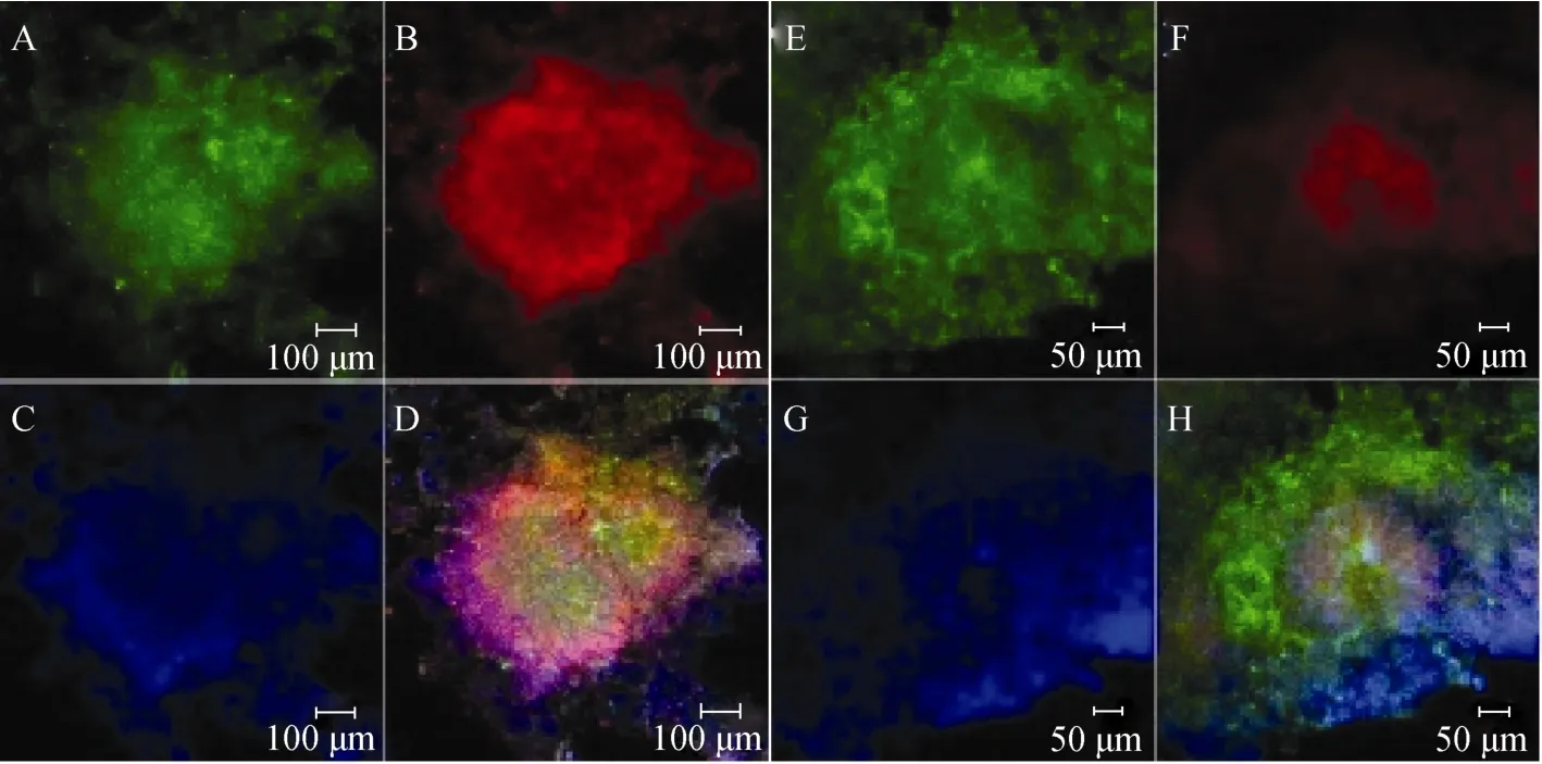
圖1 LYON-ES細胞在神經分化第12天分化為神經干細胞Fig.1 LYON-ES cells differentiated into neural stem cells at the 12th day
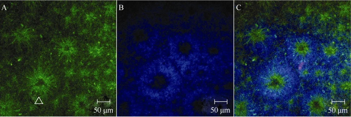
圖2 Rosettes結構神經干細胞經(RNSCs)bFGF/EGF擴增Fig.2 Rosettes neural stem cells(RNSCs) proliferate with bFGF/EGF
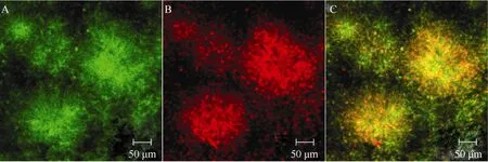
圖3 手工挑選Rosettes結構神經干細胞經bFGF/EGF擴增傳代培養Fig.3 The Rosettes neural stem cells proliferation and differentiation with bFGF and EGF
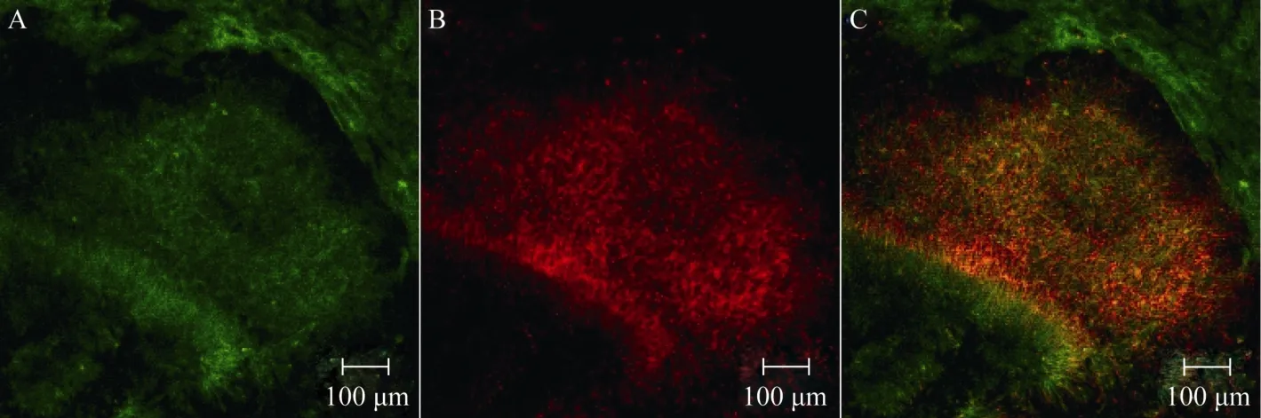
圖4 手工挑選Rosettes結構神經干細胞經擴增傳代培養(無bFGF/EGF)Fig.4 The Rosettes neural stem cells proliferation and differentiation without bFGF and EGF
ESCs具有分化為身體的所有類型細胞的能力。長期以來, ESCs向神經細胞的分化一直是研究熱點。目前, 體外定向誘導ESCs向神經分化的方法多樣, 依據分化原理可以分為以下幾種:化學物質誘導法(Bain et al, 1995)、EB形成法(Reubinoff et al, 2001; Kuo et al, 2003; Itsykson et al, 2005)、共培養或條件培養基誘導法(Takagi et al, 2005;Yan et al, 2005)、基因修飾法和單層培養誘導分化法Nestin (Andressen et al, 2001)、Sox2(Li et al, 1998)、Nurr1(Kim et al, 2002)、bHLH(Kanda et al, 2004)等。其中EB形成法又稱譜系選擇法, 是目前胚胎干細胞向神經細胞分化研究中最常用的方法。該分化方法具有三維結構, 可以模擬早期胚胎發育的許多特點, 廣泛應用在靈長類神經分化研究中(Reubinoff Et al, 2001; Kuo et al, 2003; Itsykson et al, 2005)。本實驗參照Pankratz et al ( 2007)建立的綜合EB法和單層法優點的分化體系, 建立了穩定高效的神經干細胞分化體系, 該體系分化采用無血清培養基, 得到了Nestin和Pax6高表達的神經干細胞。
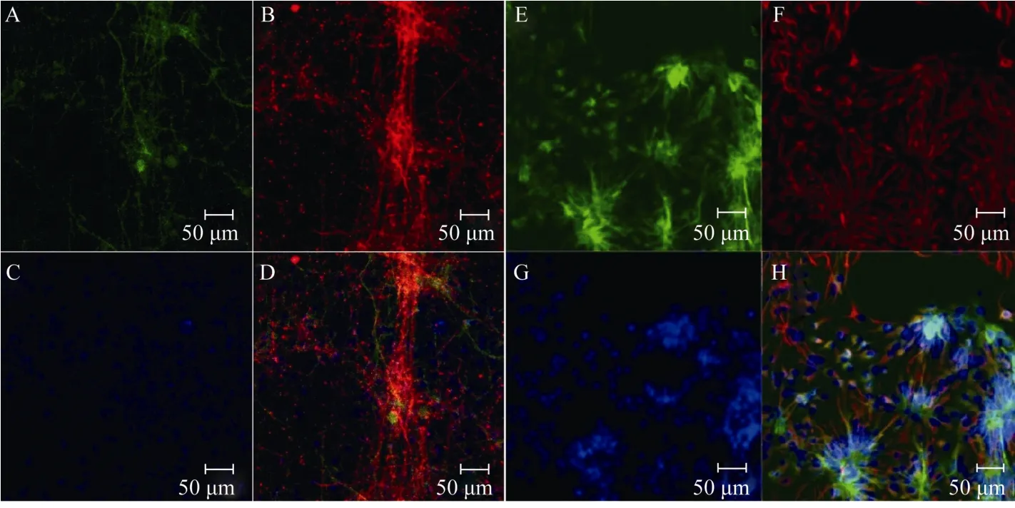
圖5 經bFGF/EGF擴增后的Rosettes神經干細胞向神經細胞分化Fig.5 The Rosettes neural stem cells proliferate with bFGF/EGF and differentiate into neurons and glia cells

圖6 經bFGF/EGF擴增后的Rosettes神經干細胞獼猴腦內移植Fig.6 The rosettes neural stem cells proliferated by adding bFGF/EGF were transplanted into rhesus monkey brain
Rosettes結構的NSCs(R-NSCs)具有廣泛的發育潛能。因此, 如何擴增和維持R-NSCs是開展神經疾病細胞替代性治療研究的關鍵。Li et al(2008)發現肝細胞生長因子(hepatocyte growth factor, HGF)能協同bFGF促進獼猴胚胎干細胞來源的神經前體細胞的增殖。雖然bFGF和EGF是常用的擴增和維持NSCs的關鍵生長因子(Murphy et al, 1990 ; Studer et al, 1998), 可有些學者認為, 當R-NSCs在bFGF/ EGF存在時, 會引起Rosettes結構的改變從而導致其向終端分化。bFGF具有誘導喙尾(rostro-caudal)軸廣泛類型細胞分化的能力, 經過bFGF長期擴增的細胞將很難分化成特定區域的神經元(Du&Zhang, 2004)。也有學者認為, 經過bFGF/EGF擴增后依然能夠長期維持其干細胞特性(Hong et al, 2008; Koch et al, 2009)。本實驗中, 手工挑選的R-NSCs經過bFGF/EGF擴增傳代2個月后,仍然能夠較好的維持Rosettes結構特性, 表明bFGF/EGF擴增能夠較好的維持干細胞的分化特性。
當前進行的神經干細胞移植實驗研究中, 多數都通過bFGF/EGF擴增, 移植后細胞的存活、分化及遷移效果良好(Muotri et al, 2005)。由獼猴胚胎干細胞分化得到的神經干細胞在大鼠上異種移植也能夠存活并分化(Li et al, 2005)。本實驗中, 經bFGF/ EGF擴增的獼猴Rosettes結構神經干細胞經同種移植后2個月, 仍然能夠較好地存活并向神經元分化。
總之, 通過bFGF/EGF擴增的神經干細胞能夠較好地維持其干細胞特性, 移植后能夠在宿主腦內存活并向神經細胞分化, 這為將來人類神經干細胞移植提供了更具參考價值的數據。
Amar AP, Zlokovic BV, Apuzzo ML. 2003. Endovascular restorative neurosurgery: a novel concept for molecular and cellular therapy of the nervous system[J]. Neurosurgery,52(2): 402–413.
Andressen C, St?cker E, Klinz FJ, Lenka N, Hescheler J, Fleischmann B, Arnhold S, Addicks K. 2001. Nestin-specific green fluorescent protein expression in embryonic stem cell-derived neural precursor cells used for transplantation[J]. Stem Cells,19(5): 419–424.
Bain G, Kitchens D, Yao M, Huettner JE, Gottlieb DI. 1995. Embryonic stem cells express neuronal properties in vitro[J]. Dev Biol,168(2): 342–357.
Calhoun JD, Lambert NA, Mitalipova MM, Noggle SA, Lyons I, Condie BG, Stice SL. 2003. Differentiation of rhesus embryonic stem cells to neural progenitors and neurons[J]. Biochem Biophys Res Commun,306(1): 191–197.
Chen HW, Mao Y, Li B, Wei Q, Zhang J, Wang JH, Wang JH, Wang SF, Tan T, Zhang XZ, Li J, Ma YY, Ji WZ. 2008. Neural lineage development of rhesus monkey embryonic stem cells: Insight of neurogenesis and gliogenesis in vitro[J]. Cell Res,18(S1): s148.
Chen SS, Revoltella RP, Papini S, Michelini M, Fitzgerald W, Zimmerberg J, Margolis L. 2003. Multilineage differentiation of rhesus monkey embryonic stem cells in three-dimensional culture systems[J]. Stem Cells,21(3): 281–295.
Cohen S. 1962. Isolation of a mouse submaxillary gland protein accelerating incisor eruption and eyelid opening in the new-born animal[J]. J Biol Chem,237: 1555–1562.
Du ZW, Zhang SC. 2004. Neural differentiation from embryonic stem cells: which way?[J]. Stem Cell Dev,13(4): 372-381.
Elkabetz Y, Studer L. 2008. Human ESC-derived neural rosettes and neural stem cell progression[J]. Cold Spring Harb Symp Quant Biol,73: 377–387.
Gage FH. 2000. Mammalian neural stem cells[J]. Science,287(5457): 1433-1438.
Gospodarowicz D. 1975. Purification of a fibroblast growth factor from bovine pituitary[J]. J Biol Chem,250(7): 2515–2520.
Hong SH, Kang UJ, Isacson O, Kim KS. 2008. Neural precursors derived from human embryonic stem cells maintain long-term proliferation without losing the potential to differentiate into all three neural lineages, including dopaminergic neurons[J]. J Neurochem,104(2): 316–324.
Itsykson P, Ilouz N, Turetsky T, Goldstein RS, Pera MF, Fishbein I, Segal M, Reubinoff BE. 2005. Derivation of neural precursors from human embryonic stem cells in the presence of noggin[J]. Mol Cell Neurosci,30(1): 24–36.
Kanda S, Tamada Y, Yoshidome A, Hayashi I, Nishiyama T. 2004. Over-expression of bHLH genes facilitate neural formation of mouse embryonic stem (ES) cells in vitro[J]. Int J Dev Neurosci,22(3): 149–156.
Kim JH, Auerbach JM, Rodríguez-Gómez JA, Velasco I, Gavin D, Lumelsky N, Lee SH, Nguyen J, Sánchez-Pernaute R, Bankiewicz K, McKay R. 2002. Dopamine neurons derived from embryonic stem cells function in an animal model of Parkinson's disease[J]. Nature,418(6893): 50–56.
Koch P, Opitz T, Steinbeck JA, Ladewig J, Brüstle O. 2009. A rosette-type, self-renewing human ES cell-derived neural stem cell with potential for in vitro instruction and synaptic integration[J]. Proc Natl Acad Sci USA,106(9): 3225–3230.
Kuai XL, Gagliardi C, Flaat M, Bunnell BA. 2009. Differentiation of nonhuman primate embryonic stem cells along neural lineages[J]. Differentiation,77(3): 229–238.
Kuo HC, Pau KY, Yeoman RR, Mitalipov SM, Okano H, Wolf DP. 2003. Differentiation of monkey embryonic stem cells into neural lineages[J]. Biol Reprod,68(5): 1727–1735.
Li M, Pevny L, Lovell-Badge R, Smith A. 1998. Generation of purified neural precursors from embryonic stem cells by lineage selection[J]. Curr Biol,8(17): 971–974.
Li RR, Chen HW, Chen DL, Wang SF, Zhang J, Chen R, Ji WZ. 2008. Hepatocyte growth factor promotes the proliferation of the neural progenitors derived from rhesus monkey embryonic stem cells[J]. Zool Res,29(5): 518–528. (in Chinese)
Li T, Zheng J, Xie Y, Wang S, Zhang X, Li J, Jin L, Ma Y, Wolf DP, Zhou Q, Ji W. 2005. Transplantable neural progenitor populations derived from rhesus monkey embryonic stem cells[J]. Stem Cells,23(9): 1295–1303.
Modo M, Rezaie P, Heuschling P, Patel S, Male DK, Hodges H. 2003. Transplantation of neural stem cells in a rat model of stroke: assessment of short-term graft survival and acute host immunological response[J]. Brain Res,958(1): 70–82.
Muotri AR, Nakashima K, Toni N, Sandler VM, Gage FH. 2005. Development of functional human embryonic stem cell-derived neurons in mouse brain[J]. Proc Natl Acad Sci USA,102(51): 18644–18648.
Murphy M, Drago J, Bartlett PF. 1990. Fibroblast growth factor stimulates the proliferation and differentiation of neural precursor cells in vitro[J]. J Neurosci Res,25(4): 463–475.
Okita K, Ichisaka T, Yamanaka S. 2007. Generation of germline-competent induced pluripotent stem cells[J]. Nature,448(7151): 313–317.
Pankratz MT, Li XJ, Lavaute TM, Lyons EA, Chen X, Zhang SC. 2007. Directed neural differentiation of human embryonic stem cells via an obligated primitive anterior stage[J]. Stem Cells,25(6): 1511–1520.
Reubinoff BE, Itsykson P, Turetsky T, Pera MF, Reinhartz E, Itzik A, Ben-Hur T. 2001. Neural progenitors from human embryonic stem cells[J]. Nat Biotechnol,19(12): 1134–1140.
Studer L, Tabar V, Mckay R. 1998. Transplantation of expanded mesencephalic precursors leads to recovery in parkinsonian rats[J]. Nat Neurosci,1(4): 290–295.
Takagi Y, Takahashi J, Saiki H, Morizane A, Hayashi T, Kishi Y, Fukuda H, Okamoto Y, Koyanagi M, Ideguchi M, Hayashi H, Imazato T, Kawasaki H, Suemori H, Omachi S, Iida H, Itoh N, Nakatsuji N, Sasai Y, Hashimoto N. 2005. Dopaminergic neurons generated from monkey embryonic stem cells function in a Parkinson primate model[J]. J Clin Invest,115(1): 102–109.
Wianny F, Bernat A, Huissoud C, Marcy G, Markossian S, Cortay V, Giroud P, Leviel V, Kennedy H, Savatier P, Dehay C. 2008. Derivation and cloning of a novel rhesus embryonic stem cell line stably expressing tau-green fluorescent protein[J]. Stem Cells,26(6): 1444–1453.
Wolf DP, Kuo HC, Pau KY, Lester L. 2004. Progress with nonhuman primate embryonic stem cells[J]. Biol Reprod,71(6): 1766–1771.
Yan YP, Yang DL, Zarnowska ED, Du ZW, Werbel B, Valliere C, Pearce RA, Thomson JA, Zhang SC. 2005. Directed differentiation of dopaminergic neuronal subtypes from human embryonic stem cells[J]. Stem Cells,23(6): 781–790.
Rhesus monkey embryonic stem cells differentiation, proliferation and allotransplantation
DONG Jin-Run1,2,3,+, GUO Li-Yun4,+, QU Jia-Gui1,2,5, QI Ren-Li1,2,5, WANG Wen-Chao1,2, XIAO Chun-Jie3,*, WANG Zheng-Bo1,2,*
(1. State Kay Laboratory of Brain and Cognitive Science, Institute of Biophysics, the Chinese Academy of Sciences, Beijing 100101, China; 2. Kunming Institute of Zoology, the Chinese Academy of Sciences, Kunming 650223, China; 3.Life Sciences Faculty of Yunnan University, Kunming 650223, China; 4.Department of Ophthalmology, the Fourth Affiliated Hospital of Kunming Medical College, Kunming 650021,China; 5School of Life Science, University of Science and Technology of China, Hefei 230026, China)
To investigate the characteristics of rhesus monkey embryonic stem cells and to promote their clinical application, the differentiation and proliferation of rosettes neural stem cells from GFP marked rhesus monkey embryonic stem cells were studied The results showed that: 1) A stable and high-efficient neural differentiation system was established. More than 95% of the embryonic stem cells were differentiated into neural stem cells on the 12thdays of differentiation; 2) the rosettes neural stem cells differentiated from the rhesus monkey embryonic stem cells could maintain their rosettes-shape by proliferating with bFGF/EGF; 3) the neural stem cells could differentiate into neurons after transplanted into the rhesus monkey brain. In conclusion, the rosettes neural stem cells differentiated from rhesus monkey embryonic stem cells could maintain their characteristics after proliferation with bFGF/EGF and they could survive and differentiate into neurons after transplanted into the rhesus monkey brain, which strongly supports the clinical application of neural stem cells in the future.
Rhesus monkey; Neural stem cells; Rosettes; Basic fibroblast growth factor;Epidermal growth factor
董錦潤, E-mail: vivid_run@126.com;郭立云,昆明醫學院在讀博士研究生, kitteyyun@yahoo.com.cn
Q959.848;Q421; Q813.7
A
0254-5853-(2012)01-0043-06
book=48,ebook=388
10.3724/SP.J.1141.2012.01043
2011-12-01;接受日期:2011-12-22
;“973”項目(947703);國家自然科學基金項目(31070963, 30670669)
?通信作者(Corresponding authors), E-mail: wangzb@mail.kiz.ac.cn
+并列第一作者(Authors contributed equally to the work)
猜你喜歡
小獼猴智力畫刊(2022年9期)2022-11-04 02:31:42
中學生數理化·中考版(2022年11期)2022-02-16 07:01:20
哲學評論(2021年2期)2021-08-22 01:53:34
中華詩詞(2019年7期)2019-11-25 01:43:04
小哥白尼(趣味科學)(2019年6期)2019-10-10 01:01:50
模具制造(2019年3期)2019-06-06 02:10:54
影視與戲劇評論(2016年0期)2016-11-23 05:26:01
發明與創新(2016年38期)2016-08-22 03:02:52
太空探索(2016年5期)2016-07-12 15:17:55
現代企業(2015年9期)2015-02-28 18:56:50