Echocardiography in the diagnosis of Shone’s complex and analysis of the causes for missed diagnosis and misdiagnosis
2022-06-23 02:06:24YeDanLiHongMengKunJingPangMuZiLiNanXuHaoWangShouJunLiJunYan
World Journal of Clinical Cases 2022年11期
lNTRODUCTlON
Shone’s complex is a rare congenital heart disease characterized by multiple left heart obstructive defects, including coarctation of the aorta (CoA), valvular stenosis, and mitral stenosis[1-4]. Those defects interfere with the normal flow of oxygenated blood from the left heart. There are complete and incomplete forms of the syndrome, as well as possible combinations with other heart defects such as patent ductus arteriosus, interrupted aortic arch, bicuspid aortic valve, atrial septal defect, and ventricular septal defect[5]. The spectrum of symptoms, treatments, and outcomes will vary according to the number of defects[6-10]. Shone’s complex represent about 0.7% of the patients with congenital heart disease[11] or 0.03% of echocardiography examinations[12]. The long-term prognosis is poor, and the perioperative mortality rates are 24%-27%[13,14].
Echocardiography is a non-invasive imaging modality that provides hemodynamic information in a short period and at the patient bedside. It can be used to reveal abnormal left ventricular wall motions,right ventricle dilation, an intimal flap in the ascending aorta, pericardial effusion, left ventricular ejection fraction[15-19]. It is a non-invasive, rapid, inexpensive diagnostic modality for a number of heart conditions such as pericardial tamponade, acute coronary syndrome, cardiomyopathy, pulmonary embolism, and Stanford type A aortic dissection[19,20]. It can also be used for the diagnosis, follow-up,and management of congenital heart diseases[21-23].
The studies about the use of echocardiography for the diagnosis of Shone’s complex are mainly limited to case reports[5,24-26] or small case series[12,27,28]. Nevertheless, a study suggested that echocardiography is invaluable in the characterization of the left heart defects found in Shone’s complex, but that diagnosis is complicated by the high variability of the possible combinations of defects[27]. This could result in a missed diagnosis or misdiagnosis. Additional studies are necessary to determine the exact value of echocardiography in the diagnosis of Shone’s complex.
Therefore, this study aims to examine the use of echocardiography in the diagnosis of Shone’s complex and to analyze the possible causes of missed diagnosis and misdiagnosis. The results could support the use of echocardiography for the diagnosis of Shone’s complex.
In addition to providing unified technical training and consistent examination conditions, we employed skilled sonographers to minimize bias and strictly controlled objective indicators, thus facilitating diagnoses according to diagnostic criteria. The equipment included Philips IE33 and EPIQ 7C systems, with the S8-3 (3-8 MHz) and S5-1 (1-5 MHz) probes (Philips, Best, The Netherlands). If the children did not cooperate with the examination, a 0.5 mL/kg chloral hydrate solution was orally administrated for sedation. The children were in the horizontal or left lateral position, and echocardiography was performed in the order of subxiphoid, parasternal area, cardiac apex, and suprasternal fossa. The diagnosis was made using the three-segment method, paying special attention to the subxiphoid four-chamber view, parasternal left ventricular long-axis view, parasternal four-chamber view, parasternal left ventricular short-axis view, apical four/five-chamber view, and suprasternal fossa view.
MATERlALS AND METHODS
Study design and patients
Echocardiography was performed within one week before surgery. The ultrasonic examinations of all children were performed by a sonographer with more than 5 years of working experience.
The inclusion criteria were:(1) Surgically confirmed Shone’s complex; and (2) Underwent echocardiography, and qualified images were available. Patients with incomplete clinical data were excluded.
Diagnostic criteria
In 1963, Shone[29] reported the feature of Shone’s syndrome, which includes Annulo-Leaflet mitral ring (ALMR), parachute mitral valve (PMV), subaortic stenosis (subAS), and CoA. Shone’s syndrome is a rare form of congenital heart disease that consists of several heart defects, including ALMR, PMV,subAS, and CoA. The corresponding pathological changes are as follows:ALMR occurs in the septum of the region above the annulus of the mitral valve. PMV is a form of congenital mitral stenosis where the main pathological change is papillary muscle fusion, which involves mitral chordae tendineae attaching to a single dominant papillary muscle, leading to the inability of the mitral valve to fully open during ventricular diastole. There are two common types of subAS:(1) Limited subaortic stenosis includes fibromuscular septum inferior stenosis and septum inferior aortic stenosis, which is caused by 1.0 - 1.5 cm fibrous septums below the aortic valve; and (2) Diffuse subaortic stenosis is tubular stenosis caused by diffuse thickening of the outflow tract muscle in the left ventricle. Coarctation of the aorta is a local or diffuse narrowing of the aorta that results in reduced blood flow.
She made her living mostly by selling the milk of a flock of goats; but she was very, very poor, and not very strong, and often used to wonder how she would live if she got too weak or ill to attend to her goats
When only two or three of the abnormalities are present, Shone’s complex is diagnosed as the incomplete form. Delmo[30] believe that a mitral valve abnormality of the inflow tract is the main factor that affected surgical effects. Therefore, when there are outflow abnormalities complicated with ALMR or mitral stenosis, Shone’s complex can be diagnosed as the incomplete form.
Echocardiography
This was a retrospective study of patients who underwent echocardiography and repair surgery at Fuwai Hospital(Beijing,China)from February 14,2008,to November 22,2019.The study was approved by the ethics committee of Fuwai Hospital, Beijing, China (2016YFC1302000). The requirement for informed consent was waived by the committee because of the retrospective study nature.
If you are tired of travelling before you come to the Land of Immortality, open this box and look at my picture, and you will be borne along either on earth or in the air, quick as thought, or swift as the whirlwind
Surgery
The patients underwent repair surgeries according to different combinations of defects under general anesthesia, including mitral valvuloplasty, resection of supravalvular septum, patch angioplasty for CoA, and CoA resection and end-to-end anastomosis.
Follow-up
The patients were followed once a year at the outpatient clinic after surgery. Echocardiography was performed to observe the forward flow velocity and regurgitation of the mitral valve, forward flow velocity and regurgitation of the aortic valve, and the descending aortic flow velocity, and determine the presence or absence of postoperative re-obstruction (defined as descending aortic flow velocity of <2 m/s).
Statistical analysis
Only descriptive statistics were used. Age was presented as median (range), and categorical variables were presented as prequencies and percentages.
The statistical methods of this study were reviewed by Ye-Dan Li, Kun-Jing Pang, Mu-Zi Li, Nan Xu,Hao Wang, Shou-Jun Li and Jun Yan from State Key Laboratory of Cardiovascular Disease, Fuwai Hospital, National Center for Cardiovascular Diseases, Chinese Academy of Medical Sciences and Peking Union Medical College.
RESULTS
Characteristics of the patients
The characteristics of the 66 patients are shown in Table 1. The patients were 2.7 (0.8-5.6) years of age,and 54.5% (36/66) were male. Twenty (30.3%) were born by cesarean section, and 10 (15.2%) had a history of heart surgery. The most common heart defect was an ALMR (50/66, 75.8%), followed by CoA(43/66, 65.2%). None of the patients showed signs of cyanosis, while only one patient displayedsymptoms of dyspnea and left heart failure (1/66, 1.5%). The patients had a variety of combinations of defects (Table 1). Only two (3.0%) patients had all four defects. None of the patients had a family history of congenital heart disease.

Missed diagnoses
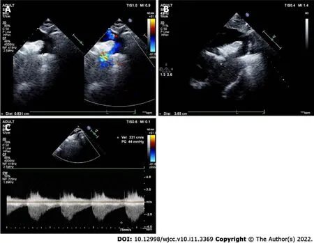
The preoperative echocardiographic findings were examined against the intraoperative findings.Echocardiography missed an ALMR in 31 patients (47.0%), a PMV in one patient (1.5%), subaortic stenosis in one patient (1.5%), and CoA in two patients (3.0%). Figures 1-4 present typical echocardiography images of Shone’s complex.
A little old man and a little old woman: While we are first introduced to the old couple in the story, this tale is not really theirs, but that of the gingerbread man


DlSCUSSlON
Shone’s complex is a rare syndrome characterized by congenital left heart defects that can differ among the patients. This retrospective study aims to examine the use of echocardiography in the diagnosis of Shone’s complex and to analyze the possible causes of missed diagnosis and misdiagnosis. The results suggest that echocardiography is an effective, non-invasive, and low-cost method to diagnose the heart defects of Shone’s complex. Due to this disease’s complexity and interindividual variability, missed diagnosis and misdiagnosis can occur. Combining the results of echocardiography, computed tomography, and/or magnetic resonance imaging might be helpful.
Some case reports examined the use of echocardiography in some patients[5,24-26], and small case series are available[12,27,28]. Ma[27] reported 38 patients with Shone’s complex that were evaluated by echocardiography. They reported a wide variety of combinations of defects among their patients, as in the present study, and concluded that echocardiography is important in the diagnosis of Shone’s complex, but they did not examine the misdiagnoses. Kumar[28] reported five patients with Shone’s complex and transesophageal echocardiographic evaluation and highlighted the usefulness of transesophageal echocardiography. Zucker[12] suggested that ultrasound is crucial to discriminate between Shone’s complex and hypoplastic left ventricle, influencing the physician’s management.
Turning to ask the Beast what it could all mean, Beauty found that he had disappeared, and in his place stood her long-loved Prince! At the same moment the wheels of a chariot were heard upon the terrace, and two ladies entered the room
Shone’s complex is a rare syndrome characterized by congenital left heart defects that can differ among the patients.
Subaortic stenosis was misdiagnosed as aortic stenosis in one case. Because the subaortic septum is often very close to the aortic valve, subaortic stenosis caused by the subaortic septum is commonly mistaken for aortic stenosis. Therefore, it is easy to misdiagnose the condition if the clinician lacks experience or does not make careful observations.
? #p# Ye--e--s! Is it you that has taken my dove? Ye--e--s! Was it you that made me kill my eleven daughters? Ye--e--s! Are you coming back again? That may be, said Esben
ONCE upon a time there lived in a certain village a little country girl, the prettiest creature who was ever seen. Her mother was excessively fond of her; and her grandmother doted on her still more. This good woman had a little red1 riding hood1 made for her. It suited the girl so extremely well that everybody called her Little Red Riding Hood2.
In addition, 10 children in this study had a history of heart surgery, but misdiagnosis or missed diagnosis still occurred because previous surgical procedures were also planned based on the results of echocardiography. For example, if only a mitral valve defect was found and CoA was missed at that time, only the mitral valve was treated during surgery, and the CoA was still missed.
The authors thank all the medical workers at the Ultrasound Department of Fuwai Hospital, Chinese Academy of Medical Sciences, for their help in this study.
Patients were treated with torasemide tablets and potassium citrate granules after surgery. Surgical methods of inflow tract obstruction mainly included ALMR removal, chordae tendineae release,papillary muscle incision, and mitral valve replacement. Approaches for outflow tract obstruction primarily involved aortic coarctation resection, end-to-end anastomosis, subvalvular septum removal,Among the 66 patients, seven underwent secondary surgery. Besides, there were four cases of complete Shone’s syndrome and 62 cases of incomplete Shone’s syndrome.
This study has limitations. Most previous studies are either case reports or small case series, and the present study is probably the largest series so far, with= 66, but it is still a small series to draw firm conclusions. All patients were from a single center, and future studies should include multiple hospitals.Indeed, the disease is rare, and collective research efforts should be undertaken. Finally, the available data were limited to those available in the charts.
CONCLUSlON
In conclusion, echocardiography is an effective, non-invasive, and low-cost method to diagnose the heart defects of Shone’s complex. Due to the complexity and interindividual variability of the syndrome,missed diagnosis and misdiagnosis may easily occur. Future studies should examine the combination of multiple imaging modalities, including echocardiography, computed tomography, and magnetic resonance imaging.
ARTlCLE HlGHLlGHTS
Research background
An innovation of the present study is the validation of the preoperative echographic findings with the intraoperative findings. A surprising result is that echocardiography missed an ALMR in 47.0% of the patients or 62.0% of the patients with an ALMR, while PMV (1.5% of the patients or 5.0% of the PMVs),subaortic stenosis (1.5% of the patients or 4.0% of the subaortic stenoses), and CoA (3.0% of the patients or 4.7% of the CoAs) were missed in smaller proportions of patients. Various reasons might be involved.The mitral ring is very small. Sometimes, only the ridge adhered to the mitral valve, or only to the anterior and posterior leaflets or annulus of the mitral valve, or did not adhere to the mitral valve but was very close to it. In these cases, it was difficult to identify an ALMR on echocardiography. The mitral valve leaflets can also be thickened and enhancing the echo, which can easily cover the supravalvular ring on the images. If the sonographer is inexperienced, the ALMR might be missed without further careful observation when PMV and mitral stenosis were found. Regarding PMV. If the left ventricular short-axis papillary muscle is not carefully checked, the anomaly of the papillary muscle can be missed.The flow velocity can be increased in the presence of mitral stenosis. If the sonographer considered the increase in flow velocity as a result of mitral stenosis, PMV might be missed due to not paying further attention. Subaortic stenosis is classified as the membranous type and fibromuscular type (isolated and diffuse stenosis). Usually, the manifestation of fibromuscular stenosis is obvious, and it cannot be missed. On the other hand, the membranous type is easy to be missed, because the subvalvular septum is sometimes very small, or the septum is close to the aortic valve. CoA can be classified as two types according to the different positions of the arterial duct:preductual and postductal. If the color Doppler and spectral Doppler images of the descending aortic arch on the suprasternal fossa view are not carefully observed, CoA can be missed. CoA patients are often accompanied by post-stenotic dilation of the descending aorta, which may suggest CoA. When children develop left ventricular wall hypertrophy or decreased left ventricular systolic function, the presence of CoA can be considered. In addition, many patients with anomalies of the bicuspid aortic valve have CoA and should be carefully screened.
The coffee shop, with its excellent location and coffee, had helped make his vacation a pleasant one. But he knew in his heart, had his brother been there to join him just one day, his vacation would have been a perfect one.
Research motivation
To use echocardiography in the diagnosis of Shone’s complex and analyze the causes of missed diagnosis and misdiagnosis.
Research objectives
Sixty-six patients were included.
Research methods
This was a retrospective study of patients who underwent echocardiography and repair surgery from February 14, 2008, to November 22, 2019. The patients were followed once a year at the outpatient clinic after surgery.
Research results
Sixty-six patients were included. The patients were 2.7 (0.8-5.6) years of age, and 54.5% were male. Ten(15.2%) had a history of heart surgery. The most common heart defect was the Annulo-Leaflet mitral ring (ALMR) (50/66, 75.8%), followed by coarctation of the aorta (CoA) (43/66, 65.2%).
Research conclusions
Echocardiography is an effective method to diagnose the Shone’s complex. Due to this disease’s complexity and interindividual variability, Improving the understanding of the disease can reduce misdiagnosis and missed diagnosis.
Research perspectives
This was a retrospective study with the largest sample size which aimed to examine the use of echocardiography in the diagnosis of Shone’s complex and to analyze the possible causes of missed diagnosis and misdiagnosis. Sixty-six patients were included. The preoperative echocardiographic findings were examined against the intraoperative findings. Echocardiography missed an ALMR in 31 patients, a parachute mitral valve in one patient, subaortic stenosis in one patient, and CoA in two patients. Due to this disease’s complexity and interindividual variability, echocardiography missed diagnosis can occur.Combining the results of echocardiography, computed tomography, magnetic resonance imaging might be helpful.
There was once a fisherman and his wife who lived together in a little hut close to the sea, and the fisherman used to go down every day to fish; and he would fish and fish
And what, said the tiger-lily? “Hark, do you hear the drum?— ‘turn, turn,’—there are only two notes, always, ‘turn, turn.’ Listen to the women’s song of mourning! Hear the cry of the priest! In her long red robe stands the Hindoo widow by the funeral pile. The flames rise around her as she places herself on the dead body of her husband; but the Hindoo woman is thinking of the living one in that circle; of him, her son, who lighted those flames. Those shining eyes trouble her heart more painfully than the flames which will soon consume her body to ashes. Can the fire of the heart be extinguished in the flames of the funeral pile?”
But the Prince begged and implored3 so long, that at last his father consented to let him go, and furnished him with gold and silver as he had done his brothers
The prognosis of patients after surgery was as follows:one patient developed a third-degree atrioventricular block and had a permanent pacemaker installed. Another case had cyanosis and dyspnea and underwent mitral and tricuspid valve repair. One patient had severe mitral insufficiency in the early stage and received a mechanical mitral valve replacement three days after the operation.Besides, one case underwent ALMR resection nine years after the first operation. There were no instances of in-hospital deaths.
When her hunger was satisfied, the old witch, growing drowsy29, lay down on the stove and said: Listen to me well, and do what I bid thee. Tomorrow when I drive away, do thou clean the yard, sweep the floors and cook my supper. Then take a quarter of a measure of wheat from my store house and pick out of it all the black grains and the wild peas. Mind thou dost all that I have bade; if not, thou shalt be eaten for my supper.
Li YD, Xu N and Li MZ carried out the studies, participated in collecting data, and drafted the manuscript; Li YD, Meng H, Pang KJ and Wang H performed the statistical analysis and participated in its design; Li YD, Li SJ, and Yan J participated in acquisition, analysis, or interpretation of data and draft the manuscript; all authors read and approved the final manuscript.
The study was approved by the ethics committee of Fuwai Hospital, Beijing,China (2016YFC1302000).
The requirement for informed consent was waived by the committee because of the retrospective study nature.
Cherry-scented smoke from Grampy s pipe kept the hungry mosquitoes at bay while gray, wispy1 swirls2 danced around our heads. Now and again, he blew a smoke ring and laughed as I tried to target the hole with my finger. I, clad in a cool summer nightie, and Grampy, his sleeveless T-shirt, sat watching the traffic. We counted cars and tried to guess the color of the next one to turn the corner.
We have no financial relationships to disclose.
No additional data are available.
This article is an open-access article that was selected by an in-house editor and fully peer-reviewed by external reviewers. It is distributed in accordance with the Creative Commons Attribution NonCommercial (CC BYNC 4.0) license, which permits others to distribute, remix, adapt, build upon this work non-commercially, and license their derivative works on different terms, provided the original work is properly cited and the use is noncommercial. See:https://creativecommons.org/Licenses/by-nc/4.0/
China
Ye-Dan Li 0000-0002-3992-2972; Hong Meng 0000-0002-2028-4018; kun-Jing Pang 0000-0003-0605-2680;Mu-Zi Li 0000-0001-8997-3682; Nan Xu 0000-0003-4746-7655; Hao Wang 0000-0003-0780-248X; Shou-Jun Li 0000-0002-6878-8733; Jun Yan 0000-0002-9380-8695.
Ma YJ
A
Ma YJ
 World Journal of Clinical Cases2022年11期
- World Journal of Clinical Cases的其它文章
- Pleomorphic adenoma of the left lacrimal gland recurred and transformed into myoepithelial carcinoma after multiple operations:A case report
- Thyrotoxicosis after a massive levothyroxine ingestion:A case report
- Contrast-enhanced ultrasound manifestations of synchronous combined hepatocellular-cholangiocarcinoma and hepatocellular carcinoma:A case report
- Papillary thyroid microcarcinoma with contralateral lymphatic skip metastasis and breast cancer:A case report
- Del(5q) and inv(3) in myelodysplastic syndrome:A rare case report
- Ultrasound-guided local ethanol injection for fertility-preserving cervical pregnancy accompanied by fetal heartbeat:Two case reports