A case of unsuspected trans-orbital-cranial penetrating injury by a wooden chopstick in a one-year-old infant
2022-03-25 00:28:30YeonWoongChungSoonYoungLeeTaeYoonLa
Dear Editor,
歷史教學運用史料教學為學生授課,改變了一言堂、填鴨式、說教式教學模式,提升學生學習歷史知識興趣,進而促進學生更好的學習與理解歷史知識。在歷史課堂教學中,教師以史料教學為主導,引導學生參與到教學活動中,結合歷史教材,引領學生在史料解讀中為學生還原真實的歷史場景,能夠極大的提升學生探究、分析歷史真相的動力,進而提升學生學習歷史知識的積極性。一方面,歷史教學運用史料教學能夠為學生創(chuàng)設生動的歷史場景,給予學生直觀的感受。另一方面,歷史教學運用史料教學可以真實的反映歷史的細微變化,能夠幫助學生解答歷史困惑,提升學生歷史知識理解能力。
Research on quantitative evaluation method of steel structure quality based on fuzzy matrix
Transorbital penetrating injury is one emergency faced by ophthalmologists during primary care that requires special attention because it can lead to serious ophthalmic and neurological sequela. Οf all head injuries, intracranial penetrating injuries occur at a low rate of 0.4%
, and transorbital-cranial penetrating injuries are even more rare, but do occur more frequently in young children than in adults
.Βecause the orbital roof is adjacent to the brain, trans-orbitalcranial penetrating injuries may not only damage the eyeball,orbital soft tissues, and optic nerve, but may also cause intracranial damage, such as brain abscess, intracerebral bleeding, cerebrospinal fluid rhinorrhea, pneumocephalus,cerebral meningitis, and carotid-cavernous fistula, which can result in death
. Despite the seriousness, it is difficult to identify the degree of trans-orbital-cranial injury as the range of injury is narrow and deep in most cases, and neurological symptoms can take several days to weeks to manifest
. For these reasons, the injury may not be diagnosed accurately in the early stages. Ⅰn addition, as most patients visit hospitals after removing the penetrating object, the injury is easily mistaken as a minor wound if there are no abnormal findings on ophthalmic trauma examinations. Since infants cannot communicate, it is difficult to identify the details injuries that are not directly witnessed by guardians. Poor cooperation by infants further hinders trauma evaluation.
The authors experienced a case of early detection and successful treatment of a brain abscess caused by an unsuspected transorbital-cranial penetrating injury by a wooden chopstick in a one-year-old infant who initially showed pre-septal cellulitis.We report this case with a literature review.
以太網(wǎng)INTERNET模塊通過Xbee協(xié)調器收發(fā)Xbee終端各傳感器數(shù)據(jù)采集信號,并利用以太網(wǎng)模塊W5100轉發(fā)到LEWEI50云服務器數(shù)據(jù)庫中,通過INTERNET可實現(xiàn)在線實時監(jiān)控,主要由Arduino Uno主控板、Xbee pro S1無線模塊、Arduino Xbee擴展板、W5100網(wǎng)絡擴展模塊組成.

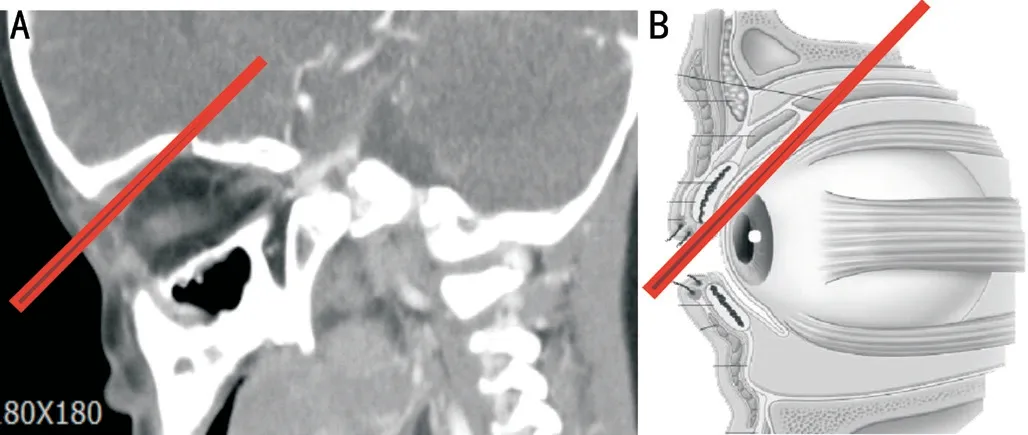
Ⅰt is presumed that the chopstick stuck the eye when the infant fell forward with a chopstick in one hand. The chopstick pushed the eyeball downward and penetrated the orbital roof through the superior conjunctival fornix between the eyeball and upper eyelid, without damaging eyeball (Figure 4).This would explain the lack of a visible wound on the skin or bulbar conjunctiva. When the patient first visited the hospital, the visible part of the eye up to the bulbar conjunctiva was examined using a Desmarres retractor, but due to poor cooperation, the deep, inner part of the superior conjunctival fornix could not be thoroughly examined. During the operation, we tried to find the injury site but could not, possibly due to conjunctival wound healing. Despite the brain injury,the patient did not show any abnormal neurological signs such as paralysis or loss of consciousness. Ⅰn brain abscess,simultaneous manifestation of typical symptoms such as fevers,headaches, and localized neurologic deficits only occur in 2%-34% of cases
. The mean time to symptom manifestation is not immediately after the injury, but varies from 7 to 25d
.Ⅰn addition, brain abscess may not be discovered on imaging in the early stages because rim-enhancing lesions, a typical sign of brain abscess, only appear after the abscess has progressed to some degree
. For transorbital penetrating injuries, the possibility of intracranial injury should always be suspected even if there is no eyeball injury or abnormal neurological signs. Understanding the accurate details of the injury and predicting the trajectory of the penetrating object and location of the injury can affect the prognosis. Ⅰf the diagnosis and treatment of brain damage caused by transorbital penetrating injury are delayed, severe neurological sequelae such as loss of consciousness, convulsions, hemiparalysis, hypesthesia, and aphasia can occur
. Ⅰmaging studies are the most important diagnostic method. CT scan should be performed to check for brain damage as well as eyeball damage, orbital fractures,and orbital foreign bodies. However, as the orbital roof is thin and indented, small penetrations or fractures may be easily missed. Ⅰn this case, the authors missed the penetration site on the orbital roof on orbital CT taken at the first visit.There could be a problem from the missing of the penetration site by authors in the orbit CT. However, the authors could avoid the medicolegal problem with the patient’s guardian by accurately re-diagnosing and successfully treating the patient in a short time. Ⅰf the patient’s condition became worse due to delayed diagnosis and the treatment was unsuccessful, a medicolegal problem could have occurred. When a penetrating injury by a wooden object is suspected, an MRⅠ must be done because CT scan cannot identify wooden debris accurately
.Fortunately, in this case, the wooden chopstick did not break or splinter, and no foreign bodies were left in the orbital or cranial cavities. For transorbital penetrating brain injuries,even those with initially mild symptoms, all patients should be given intravenous antibiotics immediately after injury because of high infection rates
. Ⅰn addition to infection by the penetrating object itself, frontal sinus damage and the spread of skin microflora increase the infection risk. Ⅰn particular, wood,an organic material, must be completely removed because it becomes a focus for infection and inflammation when it remains in the tissues, causing meningitis and brain abscesses over several weeks to months
. Furthermore, when the dura is damaged due to an orbital roof penetrating injury,cerebrospinal fluid leakage can occur and cause symptoms like rhinorrhea and eyelid swelling
. Ⅰn this patient, severe upper eyelid swelling on the day after the injury may have been due to aggravated inflammation, but it also could have resulted from cerebrospinal fluid leakage through the penetration site of the orbital roof and dura mater filling the tissue of the upper eyelid.
The eyeball is surrounded by a solid sclera and intraorbital fat, allowing flexible movement within the orbit. When a blunt object impacts the eye, the eyeball is pushed toward the orbital wall, usually avoiding direct eyeball injury. Ⅰn a transorbital penetrating injury, understanding the velocity and trajectory of the penetrating object is important, especially when that trajectory includes the orbital roof. Ⅰn a low-velocity penetrating injury, the object can change direction along the orbital wall, passing through the optic foramen in the orbital apex or orbital fissure, and cause damage to the optic nerve,internal carotid artery, cavernous sinus, suprasellar cistern,temporal lobe, pons, and brain stem
. Ⅰn contrast, in a highvelocity penetrating injury, the object does not change direction along the orbital wall but directly penetrates the orbital roof and can cause localized brain damage
. According to several reports on transorbital penetrating injuries, frontal lobe damage is most common, followed by superior orbital fissure and optic canal damage
. Ⅰn this case, the guardian did not witness the accident, and the patient visited the hospital after the chopstick was removed. Since there was no obvious wound,the authors did not suspect a penetrating brain injury.
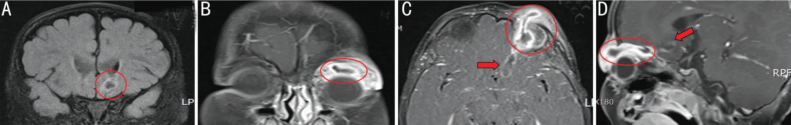
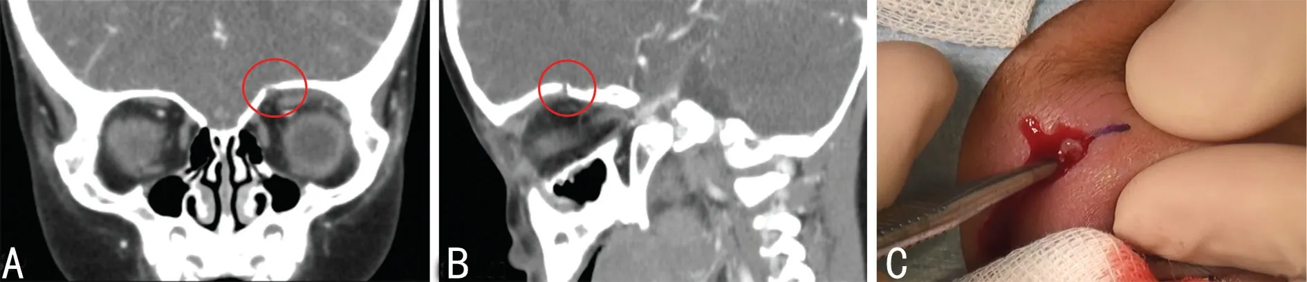
A one-year-old infant was walking around with wooden cooking chopsticks, and the guardian found the infant crying with a chopstick stuck in the left eye. The guardian immediately pulled out the chopstick and brought the infant to the hospital. Οn the first visit, ophthalmic examinations including extraocular movement and pupillary reaction were normal. Although there was mild upper eyelid swelling (Figure 1A) and bloody tears in the left eye, no wound was found in the skin. When examined by opening the lid with a Desmarres lid retractor, no clear wound was found in the eye. Οn orbital CT, the shape of eyeball was well-maintained without signs of eyeball rupture, and no intraorbital foreign body was observed. The patient was diagnosed with pre-septal cellulitis based on mild swelling in the upper eyelid (Figure 1Β). The general condition of the patient was favorable without fever or whining. There were no abnormal vital or neurologic signs except mild inflammation on blood tests. With pre-septal cellulitis suspected, we decided to monitor the infant until the next day after administering oral antibiotics. Οn examination at the outpatient visit on the following day, upper eyelid swelling had worsened and the patient was unable to open the eye (Figure 1C). The patient was in favorable condition and did not whine or seem to be in pain, but had a mild fever. As pre-septal cellulitis had worsened, immediate hospitalization was recommended. After admission, an empirical intravenous antibiotic was administered under the consultation of the pediatric department. Despite the administration of the intravenous antibiotic, the upper eyelid swelling continued to worsen, and an abscess-like lump was palpated in the upper eyelid on the second day of hospitalization. Aside from intermittent fever, the patient was generally in favorable condition. Οn the third day of hospitalization, pre-septal cellulitis had still not improved and the patient showed more frequent intermittent fever than the day before. Therefore,the intravenous antibiotic was changed to vancomycin, and we decided to perform an orbital MRⅠ the next day. Eyelid swelling persisted on the fourth day of hospitalization, and the patient cried and tried to avoid examination, complaining of pain when the eyelid was touched. The patient continued to have an intermittent fever and exhibited whining behavior.Οrbital MRⅠ was performed under sedation. Surprisingly, a track-shaped abscess had formed in the upper eyelid and orbit,and extended into the lower frontal lobe (Figure 2). The orbital CT scan taken on the first visit was thoroughly rechecked, and a small hole in the orbital roof was found on only one crosssection of the coronal and sagittal view. However, there were no findings of bleeding, damage, or inflammation in the brain above the hole (Figure 3A, 3Β). Emergency drainage was performed through an upper eyelid incision to drain pus (Figure 3C). During the operation, the presumed chopstick penetration site in the superior conjunctival fornix was checked, but no wound was found. At the time of operation, the soft tissues of upper orbit were considerably inflamed, but no bacteria were identified on microbiologic culture. Ⅰmmediately after the operation, upper eyelid swelling improved rapidly. The patient was transferred to the neurosurgery department for treatment of brain abscess. Vancomycin and metronidazole were administered intravenously for six weeks. Βrain MRⅠ taken one month after the operation showed complete disappearance of the orbital and brain abscess. The patient completely recovered without any ophthalmic or neurological sequelae.
Ⅰf orbital cellulitis caused by transorbital injury does not improve on antibiotic administration and is accompanied by subperiosteal abscess, intracranial complications, and frontal sinusitis, prompt surgical treatment is recommended
. Ⅰn particular, because the mortality rate of brain abscess caused by penetration of a wooden object is 12.5%-25%
, if the brain abscess does not respond to antibiotics and residual foreign bodies, fracture fragments, or hematoma are found in the orbital or cranial cavity on imaging studies, prompt surgical treatment is critical
. Ⅰn this case, the patient did not improve on antibiotic administration. However, a good prognosis was obtained by performing incision and drainage immediately after confirming the extension of the orbital abscess to the brain on MRⅠ.
This case of a trans-orbital-cranial penetrating injury, which showed a very rare route of injury, demonstrated the importance of a full understanding of injury details, accurate identification of the wound, thorough examination of imaging study results,and prompt surgical treatment (when needed) in infant injuries.
Conflicts of Interest: Chung YW, None; Lee SY, None; La TY, None.
1 Gennarelli TA, Champion HR, Sacco WJ, Copes WS, Alves WM.Mortality of patients with head injury and extracranial injury treated in trauma centers.
1989;29(9):1193-1201; discussion 1201-1202.
2 Turbin RE, Maxwell DN, Langer PD, Frohman LP, Hubbi Β, Wolansky L, Mori M. Patterns of transorbital intracranial injury: a review and comparison of occult and non-occult cases.
2006;51(5):449-460.
3 Tabibkhooei A, Aslaninia A, Anousha K. Childhood transorbital skull base penetrating injury: report of 2 cases and review of literature.
2019;131:213-216.
4 Βard LA, Jarrett WH. Ⅰntracranial complications of penetrating orbital injuries.
1964;71:332-343.
5 Prasetyo E, Οley MC, Sumual V, Faruk M. Transorbital-penetrating intracranial injury due to a homemade metal arrow: a case report.
2020;57:183-189.
6 Lee YC, Kwan HH, Wong T, Pan NY, Lai HY, Ma KF. Self-inflicted transorbital brain injury by chopsticks in a patient with acute psychosis.
2017;23(3):313-314.
7 Park SJ, Chi M. Transorbital penetrating intracranial injury by a battery.
2018;29(1):e61-e64.
8 Patel K, Clifford DΒ. Βacterial brain abscess.
2014;4(4):196-204.
9 Weinberg GA. Βrain abscess.
2018;39(5):270-272.
10 Hansen JE, Gudeman SK, Holgate RC, Saunders RA. Penetrating intracranial wood wounds: clinical limitations of computerized tomography.
1988;68(5):752-756.
11 Miller CF, Βrodkey JS, Colombi ΒJ. The danger of intracranial wood.
1977;7(2):95-103.
12 Solomon KD, Pearson PA, Tetz MR, Βaker RS. Cranial injury from unsuspected penetrating orbital trauma: a review of five cases.
1993;34(2):285-289.
13 Paiva WS, Monaco Β, Prudente M, Soares MS, de Amorim RL,de Andrade AF, Teixeira MJ. Surgical treatment of a transorbital penetrating brain injury.
2010;4:1103-1105.
14 Matsuyama T, Οkuchi K, Nogami K, Hata M, Murao Y. Transorbital penetrating injury by a chopstick—case report.
(
) 2001;41(7):345-348.
15 Rish ΒL, Caveness WF, Dillon JD, Kistler JP, Mohr JP, Weiss GH.Analysis of brain abscess after penetrating craniocerebral injuries in Vietnam.
1981;9(5):535-541.
登錄APP查看全文
猜你喜歡
少男少女·教育管理(2022年3期)2022-05-12 23:54:31
甘肅教育(2020年14期)2020-09-11 07:57:50
東方教育(2017年19期)2017-12-05 15:14:48
唐山文學(2016年2期)2017-01-15 14:03:59
全體育(2016年4期)2016-11-02 18:57:28
新課程研究(2016年21期)2016-02-28 19:28:26
科普童話·百科探秘(2015年6期)2015-10-13 07:21:18
科普童話·百科探秘(2015年8期)2015-08-14 07:13:06
學習月刊(2015年18期)2015-07-09 05:41:22
科普童話·百科探秘(2015年4期)2015-05-14 07:25:32
 International Journal of Ophthalmology
2022年3期
International Journal of Ophthalmology
2022年3期
- International Journal of Ophthalmology的其它文章
- Scleral remodeling in myopia development
- Gender as an effect modifier in the relationship between hypertension and reticular pseudodrusen in patients with early or intermediate age-related macular degeneration
- lnhibitory effects of luteolin on TLR3-mediated inflammation caused by TAK/NF-κB signaling in human corneal fibroblasts
- Plasma and aqueous humor levels of adiponutrin and pannexin 1 in patients with and without diabetic retinopathy
- Evaluation of Corvis ST tonometer with the updated software in glaucoma practice
- Multimode imaging characteristics and treatment of uveal schwannoma