Giant nodular fasciitis originating from the humeral periosteum:A case report
2022-03-15 11:59:28ShiLiYuPingLiSunJianLiMengJiaHongWenGao
World Journal of Clinical Cases 2022年5期
lNTRODUCTlON
Nodular fasciitis(NF)was first described as a pseudosarcomatous fasciitis by Konwaler[1]in 1955.Similar to other soft-tissue sarcomas,NF is a rapidly growing,benign proliferation of fibroblasts and myofibroblasts displaying abundant,spindle-shaped cells and high mitotic activity.NF presents most typically in the upper extremities(46%),trunk(20%),and head and neck(18%)[2].The peak incidences of NF are seen at ages 20 and 40,often presenting with tenderness,and it is a rare disease in children[3].Most NF lesions are small,measuring less than 2 cm in diameter[2,4].Periosteal fasciitis is considered a rare subtype of NF,with some case reports in the published literature and most of those were published over 20 years ago;only one case of periosteal fasciitis has been published recently,in 2017.The frequently reported sites of periosteal fasciitis are the maxilla and the hand;however,there are no reports of periosteal fasciitis in the limbs,and all reported cases described tumors that were smaller than 5 cm.
You have the sweetest voice of any who dwell here in the depths of the sea, and you believe that you will be able to charm the prince with it also, but this voice you must give to me; the best thing you possess will I have for the price of my draught
As NF has a nonspecific immunohistochemical profile[4],its histomorphological characteristics are the primary diagnostic criteria.Therefore,it remains a challenge to distinguish NF from other spindle cell lesions,particularly those of the myofibroblastic lineage.
In 2011,Erickson-Johnson[5]reported the rearrangement of thegene on chromosome 17p13 as a recurrent and specific finding in NF.Subsequently in 2013,Amary[6]foundgene rearrangements in 91% of the 34 NF cases in their study,thereby makingfluorescencehybridization(FISH)analysis a reliable and useful ancillary diagnostic test for NF.
This report presents findings from the first case of large-sized NF originating from the humeral periosteum.We emphasize the importance of highlighting this rare clinical entity,which usually represents a diagnostic dilemma.
CASE PRESENTATlON
Chief complaints
A tough mass was locally palpable on the medial side of the upper right arm and was approximately 7 cm in size.
History of present illness
Surgical tumor resection.
History of past illness
There was no history of past illness.
Personal and family history
An MRI scan showed a high signal intensity in the agglomerated pressure-fat phase near the right axillary region.The MRI images showed a lesion measuring 62 mm × 58 mm × 44 mm,with relatively well-demarcated margins.The lesion encircled the humerus,with localized thinning of the humeral cortex,and was closely related to the radial artery.
Physical examination
Intermittent pain in the right axilla for 1 mo.
1. The Bremen Town Musicians: The sources for the tale are Dorothea Viehmann and the von Haxthausen family (Zipes, Complete, 730).Return to place in story.
Laboratory examinations
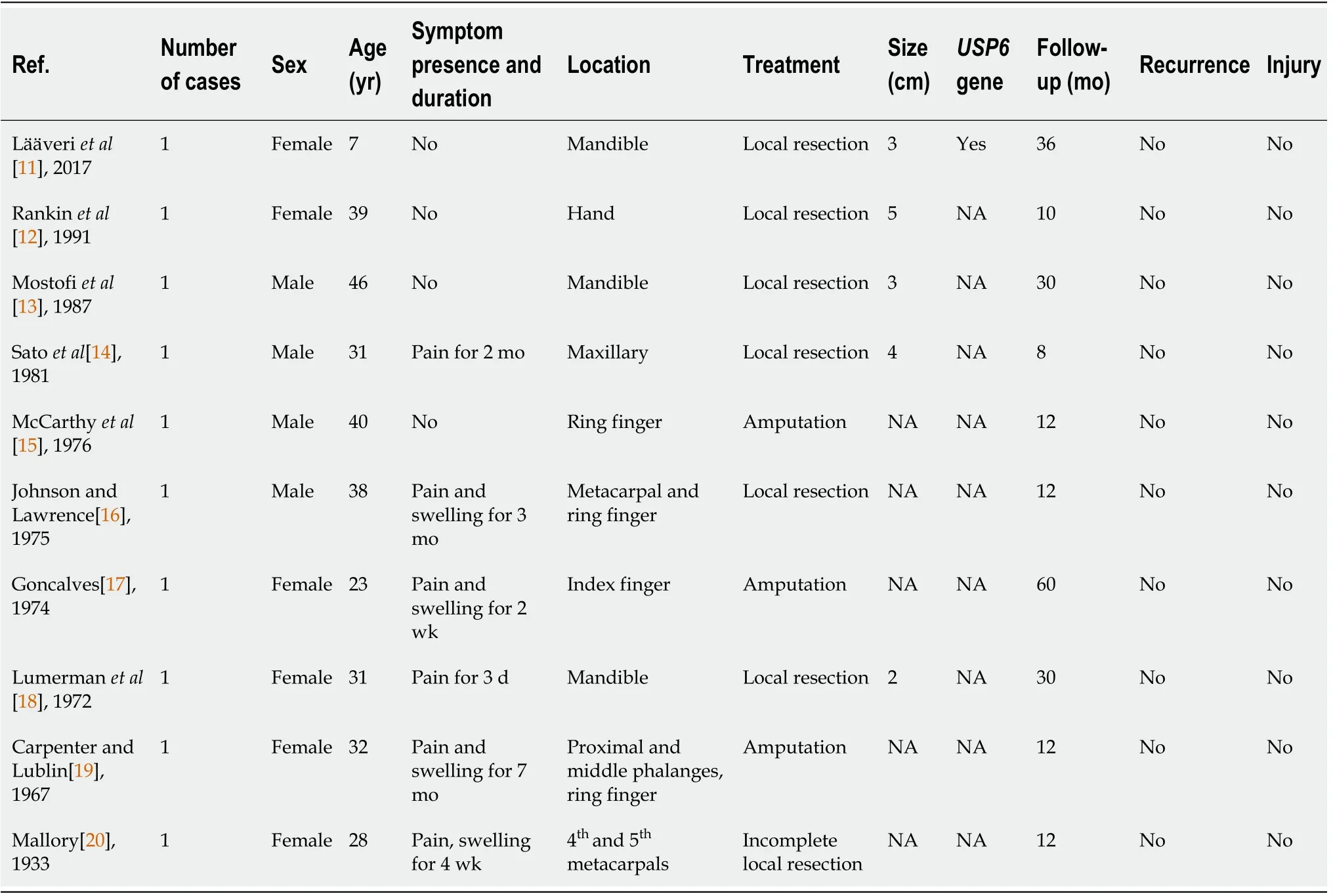
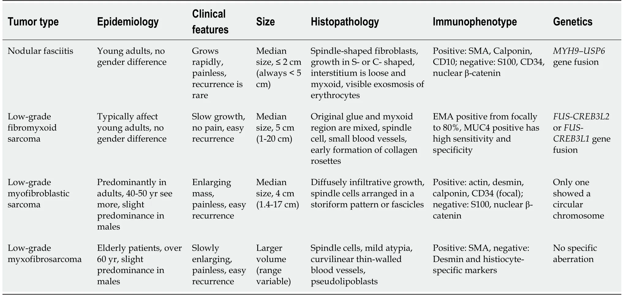
Periosteal fasciitis,a subtype of NF,is characterized by periosteal overgrowth and reactive new bone formation.There are only a few case reports(10 cases)of periosteal fasciitis in the literature,most of which were reported in the 1970s and 1980s,although one case was recently reported in 2017.Among those ten cases(four males;six females),four occurred in the jaw(one in the maxilla,three in the mandible)and six in the hand.The largest reported tumor diameter was approximately 5 cm.Most of the cases were diagnosed by histomorphological features,and FISH was undertaken in only one case in the recent literature and showedgene-related heterotopia.All patients were followed,and there are no reports of recurrence(Table 1).In our case,NF was initially diagnosed by histomorphology and immunohistochemistry;however,because of the unusually large tumor and its periosteal origin,we undertook aFISH examination.The results showed-related ectopia,which further confirmed a diagnosis of NF.The patient has shown no recurrence on follow-up for 10 mo.This report presents a rare case of clinical NF of the humeral periosteum with a tumor diameter of 7.5 cm.
Imaging examinations
There was no personal and family history.
When she came to the first jar the robber inside said softly: Is it time? Any other slave but Morgiana, on finding a man in the jar instead of the oil she wanted, would have screamed and made a noise; but she, knowing the danger her master was in, bethought herself of a plan, and answered quietly: Not yet, but presently
FlNAL DlAGNOSlS
NF.
TREATMENT
The patient had intermittent right axillary pain with no obvious cause of for 1 mo.And he found a lump under his axilla.Magnetic resonance imaging(MRI)showed a lesion measuring 62 mm × 58 mm × 44 mm,with relatively well-demarcated margins,and the lesion encircled the humerus,with localized thinning of the humeral cortex,and was closely related to the radial artery.The clinician recommended surgical treatment.
Diagnostic work-up
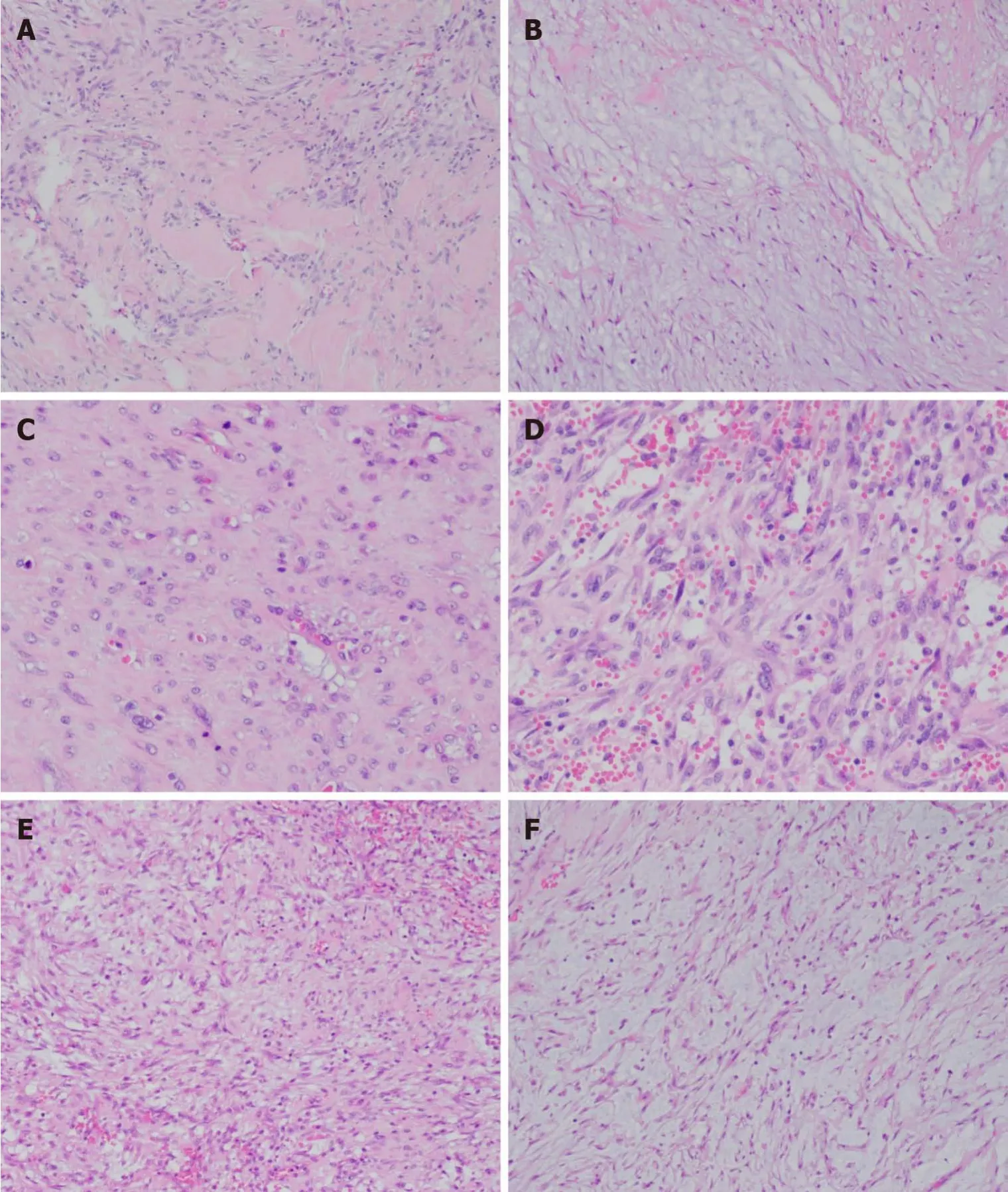
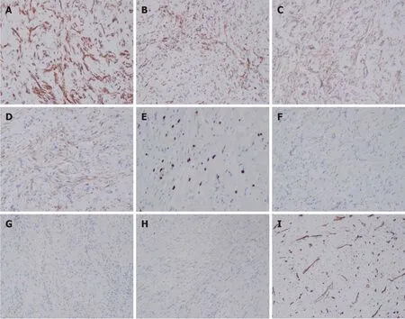
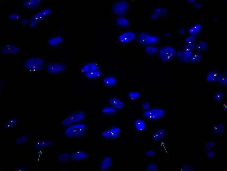
The differential diagnosis of sarcoma was made,and the patient underwent surgical tumor resection.Intraoperatively,we identified a mass with an approximate diameter of 7 cm that was closely related to the humerus,with a relatively clear boundary that separated it from the surrounding tissue.The tumor was completely separated from the periosteum.The surgical specimen was intraoperatively subjected to rapid histopathological examination.Gross examination revealed a gray nodule measuring 7.5 cm × 4 cm × 4 cm that had a reddish gray surface appearance on cross section and relatively tough texture(Figure 1).Microscopically,the lesion mainly comprised spindle-shaped fibroblast-like cells,with mucinous degeneration,mild atypia of some cells,and 3-4 mitotic figures per 10 high power fields.The intraoperative provisional pathological diagnosis was a mesenchymal neoplasm;the final diagnosis would be definitively based on the postoperative pathology.The postoperative histopathology of the lesions revealed spindle-shaped tumor cells with abundant extracellular mucoid matrix(Figure 2B and F);similarly,on examination of the frozen sections,some areas showed fibrous hyperplasia and hyaline degeneration(Figure 1A),whereas other areas had extravasation of red blood cells(Figure 2D).Tumor cells in areas with relatively high cellularity showed mild atypia(Figure 2C and D)and mitotic figures(Figure 2C).Immunohistochemistry showed that the specimen stained negative for CD34,S100,and β-catenin and positive for CD10 and SMA(Figure 3).FISH analysis revealed agene fracture rearrangement(Figure 4)with signal patterns as follows:1G1R1F 16.5%,1G1R 8.5%,2F 35.5%,1F 25.0%,1G1F 7.0%,and 1R1F 7.5%.

OUTCOME AND FOLLOW-UP
Sometimes,it may be difficult to distinguish low-grade myxofibrosarcoma from NF,especially in cases with small tumor volume and without specific immunohistochemical markers.Nonetheless,curvilinear thin-walled blood vessels and pseudolipoblasts suggest the possibility of a myxofibrosarcoma,and FISH examination shows nogene-related ectopia.
DlSCUSSlON
The published literature describes NF as a benign myofibroblastic proliferation,which was initially reported in 1955 as a pseudosarcomatous fibromatosis or fasciitis[1].The NF lesion typically develops in the subcutaneous superficial fascia of the upper limbs(46%),especially over the volar aspect of the forearm,followed by the head and neck(20%),trunk(18%),and lower extremities(16%).There are no gender differences in NF incidence,and all reported lesions measure less than 5 cm in diameter.
6. From her father she would certainly get blows: Not only is the child cold and hungry, she is also abused at home, increasing the pathos32 and stark reality of the story.Return to place in story.
No abnormalities were found in routine laboratory tests.
Due to its fast and infiltrative growth pattern,NF remains one of the most commonly misdiagnosed benign spindle cell neoplasms.A common differential diagnosis of NF is low-grade malignant myofibroblastic tumors because,despite their large size,the tumor cells are characterized by mild atypia;positive staining for actin,desmin,calponin,and CD34(focal),and negative staining for S100 and nuclear β-catenin[7-9].However,FISH shows nogene-related ectopia,and myofibroblastic tumors have a high recurrence after surgical resection.
The patient had an uneventful recovery after surgery and no further treatment was given.There was no recurrence during the 20-mo follow-up period.
Tears came to my eyes as I realized what I had been a fool to judge Al as a failure. He had not left any material possessions behind. But he had been a kind loving father, and left behind his best love.
Low-grade malignant fibromyxoid sarcoma is another differential diagnosis of NF.The identification can be comprehensively evaluated by immunohistochemical staining and molecular detection.Immunohistochemistry shows EMA positivity from focally to 80%,and MUC4 positivity has high sensitivity and specificity for the detection of fibromyxoid sarcoma[10].Molecular genetics showorgene fusion(Table 2).
Immunohistochemical staining has no specific significance in the identification of NF;however,it can be used as an auxiliary and differential diagnostic tool because spindle cells in NF often diffusely express SMA,and are negative for desmin.Recent studies have shown thathybridization has higher specificity and sensitivity in the diagnosis of NF[6],particularly in cases with uncharacteristic morphology.
Furthermore,NF can be accurately diagnosed by combining tumor morphological characteristics,immunohistochemical findings,anddetection,thereby avoiding misdiagnosis and overtreatment of patients.
Mother noticed other changes during that week, too. The children weren’t teasing or fighting as much. An argument would start and then suddenly stop for no good reason. Even Eric and Kelly seemed to be getting along better. In fact, all the children wore secret smiles and giggled17 to themselves at times.
CONCLUSlON
NF poses a diagnostic challenge as it is often mistaken for a sarcoma,or easily misdiagnosed as a sarcomatous lesion such as malignant fibrous histiocytoma or fibrosarcoma,because of its rapid growth,rich cellularity,and poorly circumscribed nature.NF is a tumor with rapid growth and relatively clear boundary,but it is sometimes difficult to distinguish from low-grade sarcoma under the microscope.When the tumor location is atypical and volume is large,the possibility of the disease should also be considered,especially during the operation,which can avoid excessive treatment.Postoperative histopathological examination of whole sections can be combined with immunohistochemical staining and,if necessary,the diagnosis can be confirmed by molecular detection.
 World Journal of Clinical Cases2022年5期
- World Journal of Clinical Cases的其它文章
- Subclavian artery stenting via ilateral radial artery access:Four case reports
- Neurothekeoma located in the hallux and axilla:Two case reports
- Diffuse invasive signet ring cell carcinoma in total colorectum caused by ulcerative colitis:A case report and review of literature
- Tacrolimus treatment for relapsing-remitting chronic inflammatory demyelinating polyradiculoneuropathy:Two case reports
- Aseptic abscess in the abdominal wall accompanied by monoclonal gammopathy simulating the local recurrence of rectal cancer:A case report
- Unusual magnetic resonance imaging findings of brain and leptomeningeal metastasis in lung adenocarcinoma:A case report