Relationship between Ki-67 and CD44 expression and microvascular formation in gastric stromal tumor tissues
2022-01-24 09:24:32BingMaXiaoTianHuangGuiJunZouWenYuHouXiaoHuiDu
World Journal of Clinical Cases 2022年2期
INTRODUCTION
A gastric stromal tumor (GST) is a type of gastrointestinal tumor.In recent years,the incidence of GSTs has been continuously increasing.Owing to the instability of their biological behavior,it is difficult to diagnose GSTs[1,2].Immunohistochemical markers can help predict the prognosis and determine the risk of GSTs.CD44 has recently been found to be an important indicator,showing specificity in many tumors.However,its expression characteristics in GSTs remain controversial[3].Ki-67,meanwhile,is involved in the process of cell proliferation and is highly expressed in breast cancer and neuroendocrine carcinoma[4].The role of neovascularization in the biological process of tumorigenesis cannot be ignored.Many studies have shown that GSTs contain large amounts of pro-angiogenic factors[5],but few studies have addressed the relationship between GSTs and microvessel density (MVD).The microvasculature plays a key role in the occurrence and development of tumors.It can also induce and mediate the biological processes underlying tumorigenesis,such as participating in the processes of metastasis and tumor invasion.MVD is thus a representative quantitative indicator reflecting tumor vascular growth.It is relevant to tumor nutrition and oxygen supply[6].This study explored the relationship between Ki-67 and CD44 expression in GST tissues and microangiogenesis.
MATERIALS AND METHODS
Tissue specimens
Tissue specimens of 86 cases of GSTs that were surgically resected in our hospital from April 2016 to February 2019 were selected for this study.The inclusion criteria were as follows:(1) Updates and interpretations of the National Comprehensive Cancer Network Clinical Practice Guidelines (2019 6version) on GSTs[7];(2) Patients were examined prior to their operationpreoperative computer tomography and gastroscopy biopsies;and (3) Patients had no history of radiotherapy,chemotherapy,or immunological treatment before surgery.This study was approved by the Medical Ethics Committee,and all baseline data of patients were complete.The criteria for exclusion were as follows:(1) GSTs were accompanied by other types of tumor diseases;(2) Data were missing and unable to be included for statistical analysis;(3)Patients had local recurrence;or (4) Pathological examinations were lacking.
Eighty-six GST patients aged 41 to 79 years,with an average of 62.0 ± 6.8 years,were selected.There were 35 males and 51 females.The lesion sites were as follows:gastric antrum (12 cases),gastric body (19 cases),and gastric fundus (55 cases).Fifty-six cases had tumors with a diameter larger than 2.0 cm;30 cases had a diameter equal to or less than 2.0 cm.There were 60 cases with mitotic counts equal to or less than five;26 cases had more than five mitotic counts.The risk classifications were as follows:very low risk (32 cases),low risk (39 cases),and medium high risk (15 cases).
His only aim was to run freely and to do it as effectively as he could. He was just being a child — just being himself—being completely in the moment. He was not looking for approval or was not worrying about whether someone was watching or not. He wasn t concerned about being judged. He didn t seem to be bothered by the fact that maybe someone would see him fall (as there were others in the park aside from him and his mother) and that it would be embarrassing if he did fall. No, all that mattered to him was to accomplish the task or activity at hand to the best of his ability. To run...and to feel the experience of running fully6 and freely. I learned a lot from that observation and experience, and have successfully brought that lesson with me in my many pursuits in life.
Immunohistochemical test
Some scholars believe the girl s active disobedience and aggression80 against a parental81 figure, albeit82 for moral reasons, are the reasons that the story has diminished in popularity.Return to place in story.
With a little kick from the inside he started the kettle off, and down the hill it rolled full tilt24; and when the fox came up, all that he saw was a large black kettle spinning over the ground at a great pace
Paraffin sections (thickness of 4 μm) were prepared in a conventional manner.The sections were de-waxed with xylene and gradient alcohol (100%,100%,95%,95%,80%,and 70%) to water,stepwise.Distilled water was used to rinse the sections twice (3 min each time),and phosphate buffered saline (PBS) was used to rinse the sections three times (3 min each time).The samples were then rinsed with tap water and soaked in distilled water for storage.Subsequently,the sections were placed in 10 mmol of LPH6.0 citrate buffer for antigen repair.Next,the sections were rinsed in a gentle manner under running water to bring them to room temperature.Primary antibodies were added to the tissues,which were then incubated for 16 h on a shaking table at 4 ℃.After incubation,the tissues were rinsed three times with PBS (5 min each time).The primary antibodies were not added to the negative group,and only PBS was added.Then,the secondary antibodies were added and incubated for 30 min before rinsing,according to the aforementioned method.One drop of DAB was then added to each section to aid in color development,following which the sections were incubated at room temperature for 5 min.The sections were then re-stained with hematoxylin and immersed in 1% hydrochloric acid alcohol for 30 s,1% ammonia alcohol for 45 s,and alcohol for 1 min.They were then transparentized with xylene and sealed with neutral gum.
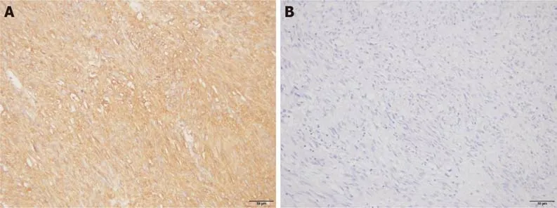
Determination of immunohistochemistry results
Positive staining of Ki-67 and CD44 proteins in the nucleus or cytoplasm is shown in yellow,brownish yellow,or brown:(1) According to the degree of staining,the results were categorized as follows:non-staining (0 points),only pale yellow staining (1 point),brownish yellow staining (2 points),and brown or black staining (3 points);and(2) According to the proportion of stained cells,the results were categorized as follows:equal to or less than 10% (one point),from 11% to 50% (two points),from 51%to 75% (three points),and more than 75% (four points).Products of staining degree and scores of positive cells that were less than three points were considered negative,whereas products that were equal to or greater than three points were considered positive.
MVD detection and counting method
Statistical analysis was performed using SPSS 21.0 software.MVD in tissues with different Ki-67 and CD44 protein expression levels is presented as mean ± SD.The two groups were compared using independent sample-tests.The positive expression rates of Ki-67 and CD44 proteins were evaluated using atest.The logistic regression model was used for multi-factor analysis.<0.05 was considered to represent a significant difference.
Statistical analysis
The segments were reviewed by two experienced pathologists with a double-blind approach.First,high MVD regions in the tissues were identified using a low-power microscope.A high-power lens with a 200-fold microscope was then used to identify individual vascular endothelial cells with brown or tan staining.The numbers of stained microvessels were counted with a microscope at five different fold magnifications,and the average value was considered the MVD (microvessels exhibit significant differences in MVD from adjacent microvessels,tumor cells,or connective tissue components).
RESULTS
Relationship between different Ki-67 protein expression levels and risk grade,mitotic counts,and GST lesion diameters
The percentages of patients with tumor risk grade (medium-to-high risk) and mitotic counts (>5) in GSTs with positive expression of the Ki-67 protein were 24.07% and 38.89%,respectively.These values were higher than those of patients with negative expression of the Ki-67 protein (6.25% and 15.63%,respectively);the difference was significant (<0.05).There were no significant differences between the positive expression rates of the Ki-67 protein in GST tissues and different lesion diameters,ages,sexes,or lesion locations (>0.05;Table 1 and Figure 1).

Relationship between different CD44 protein expression levels and risk grade,mitotic counts,and GST lesion diameters
Patients classified as medium-to-high risk with CD44 protein-positive expression in GST and patients with more than five mitotic counts accounted for 23.73% and 38.98%,respectively.These values were higher than those of Ki-67-negative patients (3.70%and 11.11%,respectively);the difference was significant (<0.05).There were no significant differences between the positive expression rates of the CD44 protein in GST tissues and different lesion diameters,ages,sexes,or lesion locations (>0.05;Table 2 and Figure 2).


Comparison of MVD in GST tissues with different Ki-67 and CD44 protein expression levels
There was no significant difference in MVD between GST tissues with positive and negative expression of the Ki-67 protein>0.05;Table 3).There was,however,a significant difference in MVD between GST tissues with positive and negative expression of the CD44 protein<0.05;Table 3).

DISCUSSION
CD44,which is a transmembrane protein belonging to the cell adhesion molecule family,theoretically plays a certain role in tumor progression and metastasis[8,9].A reduction in the expression level of CD44 would lead to poor adhesion between cells,making tumor cells more likely to shed and metastasize.However,studies have shown that the CD44 protein might play diverse and complex roles in the metastasis of different types of malignancies[10,11].In this study,an immunohistochemical technique was used to detect the expression of CD44 in GSTs.It was found that the expression of CD44 was related to the risk grade and mitotic figures of GSTs,thus indicating that mitotic figures and the primary site could be independent prognostic factors[10-12].The results of this study showed that high pathological risk grades,increased mitotic figures,and positive expression of the CD44 protein in patients with GSTs were independent risk factors for poor prognosis.CD44 could be involved in the angiogenic process in GSTs and mediate their recurrence or metastasis.Nonetheless,combining CD44 with tumor diameter and mitotic figures to more accurately evaluate and grade the risk of GSTs remains a challenge;future studies with larger sample sizes and longer follow-up times should be conducted to this effect.
In summary,the expression of Ki-67 and CD44 in GSTs has certain relationships with the tumor risk grade and mitotic changes.The expression of CD44 is related to microvessel formation in tumor tissues and the prognosis in patients with GSTs.
Reportedly,high MVD in GSTs is related to risk classification,tumor size,and mitotic counts.MVD is an independent factor that affects the prognosis in patients[13-15].The results of this study showed that there was a significant difference in MVD between tissues that were positive and negative for the CD44 protein.CD44 can promote tumor proliferation and further promote the generation of new blood vessels in tumor issues.However,new vascular basement membranes in tumors are not mature;their vascular walls are not closely arranged and are relatively loose.Thus,tumor cells can easily pass through these walls and enter the blood vessels,where they can diffuse.When the tumor spreads further,large numbers of blood vessels are further generated and MVD increases significantly.This,in turn,increases the opportunity for tumor cells to directly contact blood cells,thus promoting the infiltration and metastasis of the tumor cells.The increased expression level of CD44 provides sufficient blood supply and nutrition for angiogenesis and tumor cell proliferation.This study showed that the generation of microvessels in GSTs is relevant to the expression of CD44.
Some studies[9,16,17] have argued that increased Ki-67 expression level indicates that the tumor cells are growing rapidly,as Ki-67 can reflect the growth state of tumor cells.The results of the present study showed that there was a correlation between Ki-67 and the mitotic count.The mitotic count only reflects the M phase of cell proliferation,whereas Ki-67 is expressed in the G1,S,G2,and M phases of cell proliferation[18,19].Currently,the standard of Ki-67 expression in GSTs is unclear.This is likely because Ki-67 expression is only considered a marker of tumor proliferation from quantitative to qualitative change.In addition,the present study found that Ki-67 is more reliable than tumor size in predicting tumor risk classification and different mitotic counts.
Every morning I used to go the place where the incident happened. It was Monday morning again and the weather was bad. It was extremely6 bad! When I started to step my foot the rain started to fall. I stepped backward7 for I didn’t want to get wet. The man far behind me was wet. I got cold. I got my umbrella in my bag and started walking to the man’s direction. I was surprised the man standing is the man that I’ve been looking for! He smiled and said, “It was a year I been waiting this moment and praying someday we are going to meet again and this is it...”
That year the Fort Wayne mayor officially proclaimed16 December 21 as Amy Jo Hagadorn Day throughout the city. The mayor explained that by daring to make such a simple wish, Amy taught a universal lesson.
CD44 expression provides a certain clinical reference value for the prognoses of GSTs.Reducing MVD and inhibiting CD44 expression could suppress angiogenesis in GSTs and provide new targets for their treatment.However,the specific mechanisms need to be studied further[20].
To date,few reports have examined the relationship between Ki-67 and CD44 protein expression and the GST risk grade,as well as the changes in mitotic counts.Therefore,it is of certain significance to elucidate the mechanisms by which Ki-67 and CD44 play a role in tumorigenesis.Although there have been various speculations regarding the mechanisms of the two genes,the synergistic effects and mechanisms thereof,as well as their expression products,in the occurrence and development of GSTs remain unclear.
CONCLUSION
26. Became a queen: The young woman marries the king and becomes queen. Her ability to spin gold becomes more important than a noble birth.Return to place in story.
We found that there was no significant difference between the level of MVD in GST tissues and negative Ki-67 protein expression groups.There was no correlation between the formation of microvessels in GST tissues and the expression of the Ki-67 protein,but this lack of correlation might have been due to the limitation of the small sample size.Although Ki-67 expression was found to be irrelevant to MVD in GST tissues,it might be a candidate indicator for the prognostic evaluation of GSTs because of its association with tumor risk grade and mitotic counts.
ARTICLE HIGHLIGHTS
Research background
The incidence of gastric stromal tumors (GSTs) is increasing.The severity of a GST is often evaluated by factors such as risk classification,tumor size,and mitotic figures.However,these indicators are not very accurate.
However, she concealed19 her feelings as well as she could, and bade the intruder welcome, placing before her food and wine, hoping that when she had eaten and drunk she might take her leave
Research motivation
Few studies have addressed the relationship between GSTs and microvessel density.
Research objectives
In this study,the authors aimed to explore the relationship between Ki-67 and CD44 expression in GST tissues and microangiogenesis.
Research methods
Tissue specimens of 86 cases of GSTs were selected for this study.All cases met the inclusion and exclusion criteria.
Research results
High pathological risk grades,increased mitotic figures,and positive expression of the CD44 protein in patients with GSTs were independent risk factors for poor prognosis.
Research conclusions
The expression of Ki-67 and CD44 in GSTs has certain relationships with the tumor risk grade and mitotic changes.
Research perspectives
A deeper study with a larger sample size is needed to confirm this finding.
 World Journal of Clinical Cases2022年2期
- World Journal of Clinical Cases的其它文章
- Successful management of delirium with dexmedetomidine in a patient with haloperidol-induced neuroleptic malignant syndrome:A case report
- Using a fretsaw in treating chronic penial incarceration:A case report
- Occupational fibrotic hypersensitivity pneumonia in a halogen dishes manufacturer:A case report
- Accelerated Infliximab Induction for Severe Lower Gastrointestinal Bleeding in a Young Patient with Crohn’s Disease:A Case Report
- Tension pneumocephalus following endoscopic resection of a mediastinal thoracic spinal tumor:A case report
- Primary adrenal diffuse large B-cell lymphoma with normal adrenal cortex function:A case report