Clinical efficacy of ultrasound-guided pulsed radiofrequency combined with ganglion impar block for treatment of perineal pain
2021-04-08 08:49:56ShuiQingLiLingJiangLiGangCuiDongLinJia
World Journal of Clinical Cases 2021年9期
Shui-Qing Li, Ling Jiang, Li-Gang Cui, Dong-Lin Jia
Shui-Qing Li, Dong-Lin Jia, Department of Algology, Peking University Third Hospital, Beijing 100191, China
Ling Jiang, Li-Gang Cui, Department of Ultrasound, Peking University Third Hospital, Beijing 100191, China
Abstract BACKGROUND Ganglion impar block alone or pulsed radiofrequency alone are effective options for treating perineal pain. However, ganglion impar block combined with pulsed radiofrequency (GIB-PRF) for treating perineal pain is rare and the puncture is usually performed with X-ray or computed tomography guidance.AIM To evaluate the safety and clinical efficacy of real-time ultrasound-guided GIBPRF in treating perineal pain.METHODS Thirty patients with perineal pain were included and were treated by GIB-PRF guided by real-time ultrasound imaging between January 2015 and December 2016. Complications were recorded to observe the safety of the ultrasound-guided GIB-PRF procedure, and visual analogue scale (VAS) scores at 24 h before and after treatment and 1, 3, and 6 mo later were analyzed to evaluate clinical efficacy.RESULTS Ultrasound-guided GIB-PRF was performed successfully in all patients, and no complications occurred. Compared with pretreatment scores, the VAS scores were significantly lower (P < 0.05) at the four time points after treatment. The VAS scores at 1 and 3 mo were slightly lower than those at 24 h (P > 0.05) and were significantly lower at 6 mo after treatment (P < 0.05). There was a tendency toward lower VAS scores at 6 mo after treatment compared with those at 1 and 3 mo (P > 0.05).CONCLUSION Ultrasound-guided GIB-PRF was a safe and effective way to treat perineal pain.The 6-mo short-term clinical efficacy was favorable, but the long-term outcomes need future study.
Key Words: Ganglion impar; Perineal pain; Pulsed radiofrequency; Real-time ultrasound guidance
INTRODUCTION
Perineal pain is a common complaint, especially in women after delivery[1]. Pain receptors in this region are mainly located within the ganglion impar. Such pain is classified as sympathetic pain, and its treatments include conservative medication,physical treatment, minimally invasive treatment, psychotherapy, and surgical intervention.
Previous studies have reported that ganglion impar pulsed radiofrequency had significant pain relieving effects for refractory perineal pain and coccygeal pain[2,3].However, the puncture was often guided by X-ray or computed tomography imaging,which have limited effectiveness. The sacrococcygeal joint cannot be precisely visualized by X-ray if the patient has obvious abdominal distension. Although computed tomography can accurately show the position of the puncture needle, it inevitably increases the patient’s exposure to radiation[4,5]. In recent years, ultrasoundguided ganglion impar block was found to be an effective way to treat chronic perineal pain[6], but the patients usually needed three or more repeated blocks[7], which indicated a short duration of pain relief following a single ganglion impar block.
A previous case report observed good outcomes after the use of pudendal nerve block combined with pulsed radiofrequency to treat chronic pelvic and perineal pain[8]. However, the clinical efficacy of real-time ultrasound-guided ganglion impar block combined with pulsed radiofrequency (GIB-PRF) remain unclear. Therefore, this study aimed to evaluate the safety and clinical efficacy of real-time ultrasound-guided GIB-PRF.
MATERIALS AND METHODS
Subjects
From January 2015 to December 2016, 30 patients with perineal pain or coccygeal pain who were admitted and treated in Peking University Third Hospital were included in our study. Oral painkillers and conservative treatments were ineffective and an ultrasound-guided GIB-PRF procedure was performed. Patients with pelvic disease or histories of surgery, hip trauma, or sacrococcygeal joint fusion and calcification were excluded. All patients provided written informed consent. The study was performed in accord with the ethical principles of the Declaration of Helsinki, and was approved by the appropriate ethics committee.
Ultrasound-guided puncture
A sterile steel wire was used to assist the positioning over the skin surface. The wire was held perpendicularly to the probe and placed between the probe and skin(Figure 1A). The steel wire was moved until the “comet-tail” sign behind it with overlapping the surface of the sacrococcygeal joint (Figure 1B), which was the precise puncture point of the needle. During insertion, the lift-thrust method was used to find and trace the position of the needle tip. The needle tip was monitored using real-time ultrasound guidance until it reached the space of the sacrococcygeal joint. At that time,slight force was used to push the needle tip into the ventral sacrococcygeal joint disc,which was accompanied by an obvious sensation of fall-through. A lack of resistance was confirmed by the absence of blood, cerebrospinal fluid, or air in a syringe after injection of a small amount of saline. Anteroposterior and lateral contrast scans by a Carm X-ray unit confirmed the appropriate position of the needle tip (Figure 2).
Block and pulsed radiofrequency treatment
Sensory stimulation by an all-digital radiofrequency at 50 Hz was started to induce symptoms of perineal pain or discomfort. If the symptoms were consistent with the previous location of pain, the puncture point had reached the position of the ganglion impar. Subsequently, 3 mL of solution containing 0.2% ropivacaine and 2.5 mg diprospan was infused, followed by pulsed radiofrequency treatment for 120 s at 42oC. The above procedure was carried out collaboratively by a physician who had specialized in musculoskeletal ultrasound for over 5 years and a surgeon who had specialized in pain therapy for 15 years.
Follow-up
Visual analogue scale (VAS) scores were followed-up at 24 h before and after the treatment, and at 1, 3, and 6 mo after treatment[9]. Surgery-related complications such as rectal perforation, infection, or accidental injection of drugs into the vessels were recorded. These variables were evaluated by resident doctors in the ward or in the outpatient clinic.
Statistical analysis
Statistical analysis was performed using SPSS version 20.0 (IBM Corporation, 2011).The data were presented as the mean ± SD for continuous variables. Pairedt-tests were used to compare VAS scores before and after treatment at each of two time points.Pvalues < 0.05 were considered significant.
RESULTS
Characteristics of the study population
The mean age of these patients was 62.1 ± 12.1 years. Among the 30 patients, four were male. The mean duration of pain was 17.7 ± 9.1 mo, with a range from 6 to 36 mo.
Clinical outcomes after real-time ultrasound-guided GIB-PRF treatment
All 30 patients underwent the procedure uneventfully and without complications such as rectal perforation, infection, or accidental injection of drugs into the vessels.Compared with pretreatment VAS scores, the VAS scores significantly decreased (P<0.05) at the four evaluations performed after treatment. Compared with the VAS score at 24 h, those obtained at 1 and 3 mo after GIB-PRF treatment tended to be lower (P>0.05); the scores at 6 mo were significantly lower (P< 0.05). In addition, compared with the VAS scores at 1 and 3 mo, the scores at 6 mo after treatment demonstrated a lower tendency (P> 0.05, Table 1).
DISCUSSION
GIB-PRF is indicated in patients with perineal pain in whom oral treatment is ineffective, especially those with poor localization of pain, diffuse pain, and symptoms of a burning sensation. The ganglion impar, also called the Walther ganglion or coccygeal ganglion, is located anterior to the sacrococcygeal joint and is aretroperitoneal structure[10]. It is close to the rectum and receives sympathetic neurofibers from the sacrococcygeal area. Our study demonstrated that real-time ultrasound-guided GIB-PRF was feasible for treating perineal pain or coccygeal pain,and the key to a success treatment was related to the selection of patients and the tips for ultrasound-guided puncture.
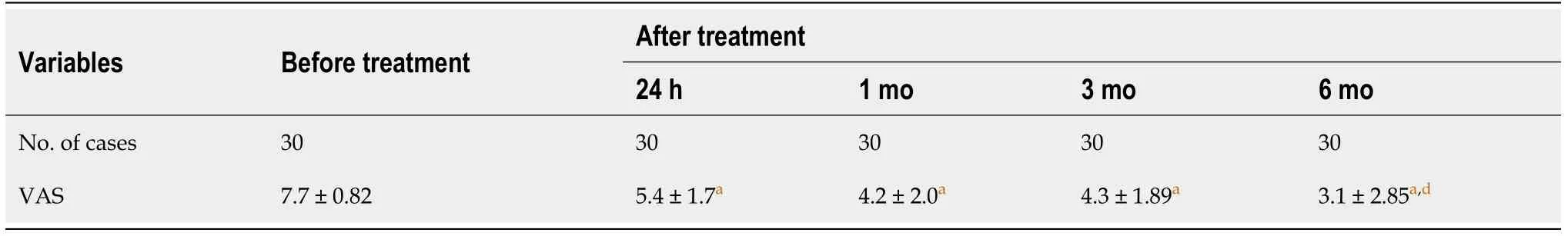
Table 1 Comparison of visual analog scale scores at different time points before and after the treatment

Figure 1 Position of the puncture point on the skin. A: A steel wire was placed perpendicular to the plane of the probe; B: The surface of the skin overlapped, causing a “comet-tail” sign behind the steel wire and the puncture position of sacrococcygeal joint (SCJ) was deeper.

Figure 2 X-ray in the lateral position. Contrast scan showing the position of the puncture needle in the sacrococcygeal joint (SCJ).
The main principle of pulsed radiofrequency treatment is the generation of a strong electromagnetic field around the tip of the electrode that relieves pain by interfering with the impulse conduction of neurons[11,12], which is one of the effective ways to treat chronic pain. The key step in the procedure is in the precise positioning of the puncture needle within the ganglion impar. X-ray or computed tomography imaging guidance was previously used for the ganglion impar puncture, but the paramedian puncture pathway, which includes the sacrococcygeal joint, coccyx, and anococcygeal ligament, varies. The reported successful puncture rate free of complications is approximately 70%, but none of the puncture methods was generally accepted as the optimal option[13]. In addition, high-frequency ultrasound is usually used to evaluate muscles, tendons, ligaments, and peripheral nerves in clinical practice, and also in the field of pain therapy[14]. Ultrasound-guided ganglion impar blocks were first reported by Guptaet al[15]in 2008 and became an effective treatment for chronic perineal pain[7,16]. However, real-time ultrasound-guided GIB-PRF for treating perineal pain was rarely studied.
Firstly, in our study, the ultrasound-guided method was roughly consistent with that reported by Johnstonet al[16]. Local, clear ultrasonograms were obtained by Johnstonet al[16], who used high-frequency probes. In our study, significant thickening of subcutaneous soft tissues in the sacrococcygeal area in obese patients prevented clear visualization of the sacrococcygeal joint because of insufficient penetration of the ultrasound waves when using a high-frequency probe. Therefore, we used a lowfrequency convex-array probe to increase penetration. The resolution of superficial structures when using a convex-array probe is relatively poor, however, visualization of the target puncture site in the sacrococcygeal joint was satisfactory, which facilitated successful insertion of the needle.
Secondly, the space within the sacrococcygeal joint is narrow and it forms an arc vertically. Thus, the in-plane view of the puncture is limited, regardless of crosssectionally or vertically scanning. Therefore, we inserted the needle out of plane,which has the advantages of space savings and a short puncture pathway. Although the needle passage cannot be displayed completely, inserting a needle perpendicular to the skin surface is easier for reaching the target, because of the relatively superficial position of the sacrococcygeal joint and the large vertical space of the articular surface.The distance between the skin and sacrococcygeal joint, as measured before insertion,was used as the reference for the depth of insertion and to avoid deep insertion.
Thirdly, another characteristic of our study was the use of a steel wire as a positioning marker. The slender steel wire was visualized on ultrasonograms as a stripe with strong echogenicity, which results from multiple interference of sound waves reflected off the metal surface, generating the “comet-tail” sign. This method greatly enhanced the accuracy of needle positioning. Therefore a careful selection of patients and tips for ultrasound-guided puncture are important to achieve a successful GIB-PRF in patients with perineal pain.
There were also several limitations in this study. Firstly, a relatively small sample size might have led to negative results, especially about post-treatment complications.In addition, this study cannot make a conclusion about superiority of block alone or pulsed radiofrequency alonevscombination of these two treatments, or ultrasound guidancevsX-ray guidance, which need further study. Lastly, a follow-up of more than 6 mo after the GIB-PRF is needed.
CONCLUSION
Ultrasound-guided GIB-PRF was a safe and effective way to treat perineal pain. The 6-mo short-term clinical efficacy was favorable. Long-term outcomes need further study.
ARTICLE HIGHLIGHTS
Research background
Despite of the efficacy of ganglion impar block alone or pulsed radiofrequency alone for treating perineal pain, the puncture is usually conducted under the guidance of Xray or computed tomography imaging. The efficacy of ganglion impar block combined with pulsed radiofrequency (GIB-PRF) for treating perineal pain remains unclear.
Research motivation
This study provides references to the clinical practices of real-time ultrasound-guided GIB-PRF in patients with perineal pain or coccygeal pain.
Research objectives
This study evaluated the safety and clinical efficacy of real-time ultrasound-guided GIB-PRF in treating perineal pain.
Research methods
Thirty patients with perineal pain who were treated by GIB-PRF with guided by realtime ultrasound imaging were analyzed. Complications were recorded to observe the safety of the treatment. VAS scores at 24 h before and after the treatment, and 1, 3, and 6 mo later were performed to evaluate clinical efficacy.
Research results
Ultrasound-guided GIB-PRF was performed successfully in all patients, and no complications occurred. Compared with pretreatment VAS scores, the VAS scores significantly decreased at the four time points after the GIB-PRF. Compared with the VAS score at 24 h after the GIB-PRF, the scores were slightly lower at 1 and 3 mo and significantly lower at 6 mo after treatment. There was a tendency toward lower VAS scores at 6 mo after GIB-PRF compared with those at 1 and 3 mo.
Research conclusions
Ultrasound-guided GIB-PRF was a safe and effective way to treat perineal pain. The 6-mo short-term clinical outcomes were favorable.
Research perspectives
The findings from this study help to establish and provide the groundwork for further studies to compare outcomes of block alone or pulsed radiofrequency alonevsthe combination of these two treatments and to investigate the long-term outcomes.
ACKNOWLEDGEMENTS
The authors would like to thank the members of Department of Pain Medicine and Ultrasound, Peking University Third Hospital.
 World Journal of Clinical Cases2021年9期
- World Journal of Clinical Cases的其它文章
- Cervical intervertebral disc degeneration and dizziness
- Contributions of aversive environmental stress to migraine chronification: Research update of migraine pathophysiology
- Expert consensus of Chinese Association for the Study of Pain on the non-opioid analgesics for chronic musculoskeletal pain
- Editorial for the special issue of the Chinese Association for the Study of Pain
- Expert consensus of Chinese Association for the Study of Pain on the radiofrequency therapy technology in the Department of Pain
- Expert consensus of the Chinese Association for the Study of Pain on pain treatment with the transdermal patch