Effect of Electroacupuncture on Platelet-derived Growth Factor and the Ultrastructure of Mitochondria in Rats with Diabetic Gastroparesis
2020-09-25 01:38:06WEIXingPENGYanZHAODongFengXIAOXiaoJuanLINYaPing
Digital Chinese Medicine 2020年1期
關鍵詞:文化
WEI Xing, PENG Yan, ZHAO Dong-Feng, XIAO Xiao-Juan, LIN Ya-Ping
The Domestic First-class Discipline Construction Project of Chinese Medicine, Hunan University of Chinese Medicine, Changsha, Hunan 410208, China
Keywords
Electroacupuncture (EA)
Diabetic gastroparesis (DGP)
Platelet-derived growth factor (PDGF)
Mitochondria
Gastric motility
ABSTRACT
Objective To observe the effect of electroacupuncture (EA)at the pressure points Zu San Li (ST36), San Yin Jiao (SP6)and Liang Men (ST21)on platelet-derived growth factor (PDGF)and the ultrastructure of mitochondria in rats with diabetic gastroparesis(DGP).
Methods Sixty Sprague Dawley (SD)rats were randomly separated into a normal control group (NC, n = 10)and a modeling group (n = 50).Rats in the modeling group received an injection of 2% streptozotocin (STZ)and a high-fat and highglucose diet for eight weeks to establish a DGP rat model.At the same time, blood glucose and a general symptom score were recorded every week.After modeling, 30 successfully modeled rats were randomly separated into the following groups: the DGP group (n = 10), the EA group (n = 10)and the metoclopramide(MP)group (n = 10).After three weeks of intervention, the gastrointestinal propulsive rate was measured by measuring the optical density (OD).The concentration of Ca2+ was determined by fluorescence immunoassay, and levels of serum insulin (INS)and PDGF were determined by ELISA.The ultrastructure of mitochondria was observed with transmission electron microscopy.
Results (1)After intervention, levels of blood glucose and the general symptom score were greatly decreased in the EA group compared to the DGP group (P < 0.01).Compared with the DGP group, the gastric emptying rate and the intestinal propulsive rate of the EA group was significantly improved (P < 0.01), and there was no statistically significant difference between the EA and the NC groups.(2)Compared with the NC group, the levels of INS in the DGP group markedly decreased (P < 0.05), but there was no significant difference of INS levels between the EA and the MP groups.(3)Compared with the DGP group, the concentration of Ca2+ in the EA and the MP groups significantly increased(P < 0.01, P < 0.05, respectively).(4)Compared with the NC group, the average OD of PDGF in the DGP group was significantly higher (P < 0.01).Compared with the DGP group,levels of PDGF in the EA group increased significantly (P < 0.01).(5)There were abundant mitochondria with a clear structure and complete cristae in the NC group.However, in the DGP group,mitochondria were severely swollen, partly vacuolated, and cristae were either fractured, absent, or shortened.In the EA group, mitochondria were slightly swollen, with clear cristae.
Conclusions Electroacupuncture at the points Zu San Li (ST36),San Yin Jiao (SP6)and Liang Men (ST21)may improve gastric motility in DGP by up-regulating the amount of PDGF and improving the ultrastructure of mitochondria.
1 Introduction
2 Materials and Methods
2.1 Animals
Sixty Sprague Dawley (SD)rats (220 - 240 g, half the number of total rats were male rats and the remaining half were female rats)were obtained from the Experimental Animal Center of Hunan University of Chinese Medicine, Laboratory animal quality certificate No.SCXK (Xiang)2009-0012.All rats were housed at the Experimental Animal Center of Hunan University of Chinese Medicine.The rats were kept at 20 - 22 °C with a relative humidity of 65% to 70% and had free access to food and sterile water.After one week of adaptive feeding, rats were included in the experiments.Ten rats were used as the normal control group (NC, half were male rats and half were female rats)and the rest were used as the modeling group to establish DGP rats.
2.2 Equipment and reagents
Streptozotocin (STZ, S0130-1G)and bovine serum album (BSA)were purchased from Sigma (Sigma,Missouri, USA).D-Hanks, type II collagenase and Dulbecco's modified eagle medium (DMEM)/F12 were from HyClone.Fluo-8 AM (CAS 1345980-40-6)was from Abcam.Metoclopramide (MP)was purchased from Shanxi Yunpeng Pharmaceutical.Sodium chloride solution (0.9%)was purchased from Hunan Kelun Pharmaceutical.Phenol red solution was from the Tianjin Guangfu Fine Chemical Research Institute.Sodium hydroxide and 20%trichloroacetic acid were purchased from Sinopharm Chemical.Glutaraldehyde (3%)was from Well-Biology.The Insulin Assay Kit (H203)and PDGF Assay Kit (H030)were from the Nanjing Jiancheng Bioengineering Institute.A blood glucose meter was purchased from Sinocare, the SDZ-II EA apparatus was from the Suzhou Medical Appliance Factory, the spectrophotometer (UV1800)was from Chengdu Shimadzu Equipment, and the inverted fluorescence microscope was from Olympus.
2.3 Experimental protocols
2.3.1 Establishment of an animal model of DGPEstablishment of rat model of DGP was based on the team’s previous researches[9,10].Except for the rats in the NC group, all other rats were modeled.After overnight fasting, but with free access to water, 50 rats in the model group were intraperitoneally injected with a single dose of 2% STZ (55 mg/kg,freshly dissolved in 0.1 mmol/L citric acid-sodium citrate buffer solution (4 °C, pH 4.5)).Seventy-two hours after injection, the development of diabetes was detected by measuring blood glucose levels from the tail vein.The rats with immediate blood glucose level of < 16.7 mmol/L were eliminated.Then, rats were irregularly fed (in morning on odd days and in afternoon on even days)with a high-glucose and high-fat diet (regular diet : lard stearin : sucrose : milk powder : egg = 58 : 15 : 20 : 5 : 2)for eight weeks and their blood glucose levels were measured every week.
In addition to the blood glucose levels, there were significant differences in the gastrointestinal propulsive parameters; for example, the condition of hair and the feces characteristics.Thirty modeled rats were randomly selected and divided into the DGP,the EA, and the MP groups.
2.3.2 Intervention methodsAll rats were fed with a normal diet during the intervention period.The location of acupuncture has been described inExperimental Acupuncture Science[11]and anatomical and anthropomorphic methods for acupuncture of rats.The pressure point Zu San Li (ST36)is located posterolateral to the knee joint and approximately 5 mm away from the capitulum fibulae.The pressure point Liang Men (ST21)or “tummy-button” is located at the junction of the upper 3/4 and lower 1/4 of the connection line between the upper edge of the sternum and the external genitalia.The pressure point San Yin Jiao (SP6)is located vertically 10 mm above the medial ankle tip of the hind legs.Rats in the EA group underwent acupuncture at Zu San Li(ST36), Liang Men (ST21)and San Yin Jiao (SP6)with an acupuncture needle (0.3 mm × 13 mm, Hwato)to a depth of 3 mm.Then, from 2 mm vertically below the locations Zu San Li (ST36), Liang Men (ST21)and San Yin Jiao (SP6)along the meridian, were respectively selected as the attachment point and the connection point of the positive pole of the EA apparatus.The SDZ-II EA apparatus was connected with points, the negative pole was connected to Zu San Li (ST36), Liang Men (ST21)and San Yin Jiao(SP6)and the positive pole was connected to the attachment points.The SDZ-II EA apparatus was conducted with a dilatational wave, 20 s, 20 Hz sparse waves alternating with 15 s, 100 Hz dense waves,20 min/d, for 15 days.The electric current was established according to the slight trembling of the skin at the attachment points.Acupuncture was conducted at the attachment points of each side, left and right sides in succession.The rats in the MP group were tied for 20 min and were treated with an intragastric administration of 1.7% MP, 1 mL/100 g,once a day, for 15 days.The NC and DGP rats were tied and treated for 20 min and were administered 0.9% sodium chloride solution by gavage (1 mL/100 g)each time, once a day, for 15 days.Blood glucose levels and the general integration of all groups were recorded.During the intervention, all rats were fed with a normal diet and allowed free access to water.After successive intervention for 15 days, all rats were starved for 12 h, and then deprived of water for 2 h.On the 16thday, rats received 2 mL phenol red solution (50 mg/dL)by gavage.Twenty minutes after receiving phenol red solution, the rats were anesthetised and specimens were collected.
2.3.3 Specimen collectionThe abdominal cavity of the rats was opened and blood was collected from the abdominal aorta.After that, the stomach was cut and the vascular and gastric serosal layers were removed,and the stomach was cut along the greater curvature.Then, the stomach was washed with 0.9% saline to collect a volume of 20 mL of the stomach contents,and the gastric emptying rate and intestinal propulsive ratio were measured.Afterwards, the stomach was quickly separated into roughly 1 mm ×1 mm × 1 mm squares of gastric antrum tissue, which were kept at ? 80 °C.
佛像是伴隨著佛教的傳播而進入的,是佛教三寶之一,隨各個時期以及不同地域的文化習俗而演變。以善法引導眾生,歷經數千年深入人心,留下了大量優秀的佛教藝術作品,在中國美術史上占據了極其重要的地位。作為豐厚文化和精神財富的代表,記載著歷代佛像作者的心力與恒定信念。
2.3.4 Criteria of the general symptom scoreThe mental state, behavioral activity, fur color and stool characteristics of the rats were observed every week,and the symptom scores were calculated with the general situation rating scale[12], as shown in Table 1.
2.3.5 Gastric emptying rate and intestinal propulsive ratioAfter deprivation of food for 12 h, rats wereadministered 2 mL phenol red solution (50 mg/dL).Twenty minutes later and after anesthesia, rats were sacrificed, followed by measurement of the total length of the small intestine.The furthest distance where sodium hydroxide (0.5 mol/L)made the small intestine turn purple served as the propelling distance of the phenol red in the intestine.The intestinal propulsive rate was calculated as (the propelling length of the phenol red/the total length of small intestine)× 100%.The whole stomach was carefully removed, then cut along the greater curvature, followed by washing with 20 mL 0.9%sodium chloride solution.Next, 20 mL sodium hydroxide(0.5 mol/L)was poured into the washed contents.One hour later, 5 mL supernatant was transferred to a centrifuge tube, and then 0.5 mL 20% trichloroacetic acid was added to the tube.The optical density (OD)of the supernatant was measured at an absorbance of 560 nm with a spectrophotometer after centrifugation(3500 r/min, 10 min).Standard phenol red solution was prepared by mixing 2 mL phenol red solution(50 mg/dL)with 18 mL distilled water, 20 mL sodium hydroxide (0.5 mol/L), and 4 mL 20% trichloroacetic acid and then the absorbance was measured.The rate of gastric emptying was calculated according to the following formula: gastric emptying (%)= (1 ?X/Y)× 100%, where X represented the obsorbance of the phenol red collected from the stomach of the animals sacrificed 20 min after the test meal and Y represented the OD of the standard phenol red solution.
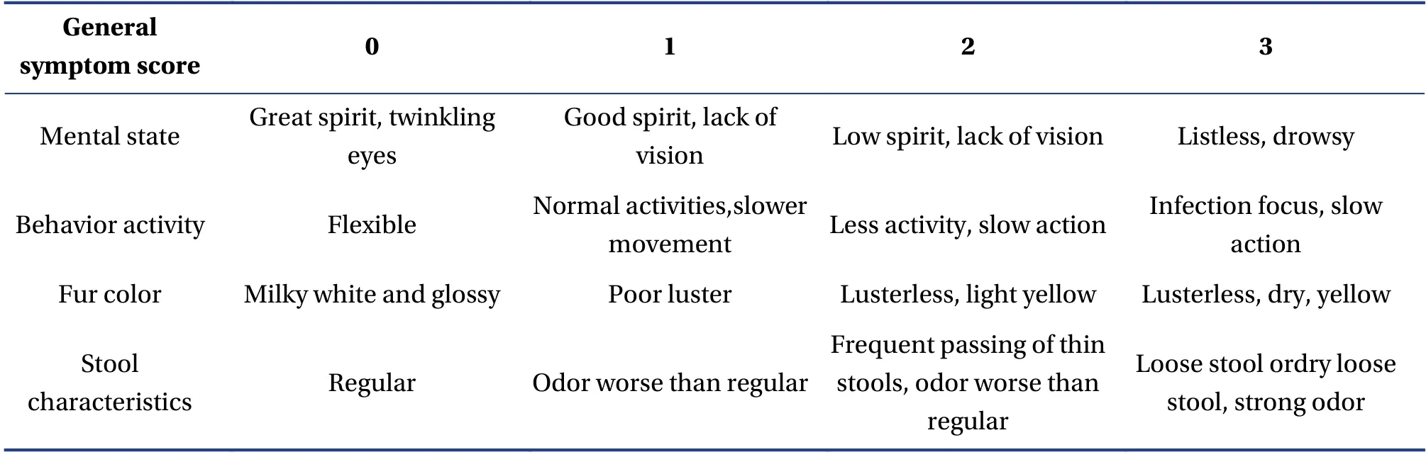
Table1 Criteria of symptom score
2.3.6 The concentration of calcium (Ca2+)To determine the concentration of Ca2+, after rinsing twice with D-Hanks solution (4 °C), the tissue was cut into 1 mm × 1 mm × 1 mm pieces and treated with type II collagenase solution, 3 - 4 times the volume of tissue, for 60 min at 37 °C.The enzymatic digestion was terminated by adding an equal volume of DMEM/F12 containing 10% FBS.The solution was centrifuged at 1000 r/min for 5 min, the supernatant was discarded, and the pellets were resuspended and passed through a cell strainer (bore diameter:0.22 μm).The cell concentration was adjusted to 1 ×10⁵/cm2.The cells were inoculated into a 96-well plate with DMEM/F12, and then put into the incubator overnight under the conditions of 37 °C, 5%CO2.When the media was removed, 4 μm Fluo-8 AM dissolved in 100 μL Hank's buffer with Hepes was added to the plate and the plate was incubated at 37 °C, 5% CO2for 1 h.The final concentration was adjusted to 4 μM and the tubes were shaken slightly.The tubes were wrapped in black paper, incubated at a 37 °C for 40 min while the tubes were gently shaken to accelerate the entry of Fluo-8 AM into the cells.After incubation, the tube was centrifuged at 1500 r for 6 min and the supernatant was discarded.At last,cells were observed and taken photos under a fluorescence microscope.
2.3.7 The concentration of INSBlood was collected and allowed to settle at 25 °C for 1 h.The blood was centrifuged at 4 °C at 2 500 rpm for 10 min.The supernatant was collected and the concentration of INS was detected by ELISA, according to the instructions of the kit from the Nanjing Jiancheng Bioengineering Institute.
2.3.8 The amount of PDGFAfter the rats were anesthetized and sacrificed, the gastric antrum tissue was quickly collected and stored in the ? 80 °C for further testing.The levels of PDGF were measured by ELISA according to the instructions of the kit from the Nanjing Jiancheng Bioengineering Institute.
2.3.9 The ultrastructure of the mitochondriaWhen specimens were obtained, the tissues were quickly put into a fixation solution for electron microscopy at 4 °C for 2 - 4 h.Samples were rinsed three times with 0.1 M phosphate buffer, each time for 15 min.Then,they were fixed in 1% osmium - 0.1 M phosphate buffer (pH 7.4)at room temperature (20 °C)for 2 h.Specimens were rinsed three times with phosphate buffer, each time for 15 min.The tissues were dehydrated sequentially with an ethanol series (50% -70% - 80% - 90% - 100% alcohol - 100% acetone -100% acetone), for 15 min each.The samples were sliced after infiltration and embedding, and dried overnight at room temperature.The tissues were observed with transmission electron microscopy(TEM)and images were collected and analyzed.
2.4 Statistical analysis
The experiment was performed using a completely randomised design, and all data were processed by the SPSS 24.0 software.Data have been expressed as the mean ± SD.Differences among groups were tested with one-way analysis of variance (ANOVA)followed by the Dunnett’s test.P< 0.05 was considered to be a statistically significant difference.
3 Results
3.1 Blood glucose levels
The comparison of blood glucose levels before modeling, after modeling and after intervention in each group has been shown in Figure 1.Before modeling, all groups were at baseline.After modeling, except the NC group, the blood glucose levels of the other groups were found to be significantly increased compared to that in the NC group (P< 0.01).Compared with the DGP group, the blood glucose levels of the EA group markedly declined to 19.5% after intervention (P< 0.01).Though EA therapy was effective, there was a significant difference when compared to the therapy in the NC group (P< 0.01).
3.2 General symptom score
The symptom scores were calculated according to the criteria in Table 2 (criteria of the general symptom score).After modeling, compared with the NC group, the general symptom score of the three groups dramatically increased (P< 0.01).When the intervention was completed, scores of both the EA and the MP groups decreased significantly (P< 0.01,P< 0.01, respectively).While, compared with the MP group, the score of the EA group decreased more significantly (P< 0.05).This indicates that EA therapy significantly decreased the systemic symptoms of DGP.
3.3 Gastrointestinal propulsive rate
Compared with the NC group, the gastric emptying rate of the DGP group showed a significant reduction(P< 0.01), indicating the DGP model had been successfully established.Compared with the NC group, the gastric emptying rate of the MP group markedly decreased (P< 0.05).And there was no obvious difference between the NC and the EA groups(P> 0.05).Compared with the DGP group, the gastric emptying rate of the EA and the MP groups significantly increased (P< 0.01,P< 0.01,respectively).
Compared with the NC group, the intestinal propulsive rate of the DGP and MP groups was found to be reduced (P< 0.01,P< 0.01, respectively).And there was no obvious difference between the NC and the EA groups (P> 0.05).Compared with the DGP group, there was a significant increase in the intestinal propulsive rate of both the EA and the MP groups(P< 0.01,P< 0.01, respectively).See Figure 2.
3.4 The amount of INS
Compared with the NC group, the amount of INS significantly decreased in the DGP group (P< 0.05).Compared with the DGP group, the amount of INS significantly increased in the EA and the MP groups(P< 0.05,P< 0.05, respectively).The amount of INS in the EA and the MP groups was comparable(P> 0.05)(Figure 3).
Table2 General symptom score ( ± s)
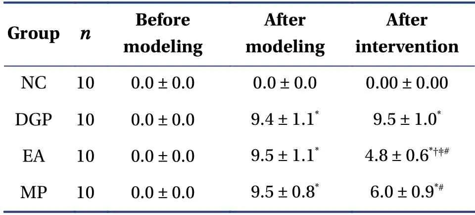
Table2 General symptom score ( ± s)
Values have been presented as the mean ± SD.*P < 0.01 compared with the NC group,?P < 0.01 compared with the DGP group,?P < 0.05 compared with the MP group at the same time point,#P < 0.01 compared with own group after modeling.
After intervention NC 10 0.0 ± 0.0 0.0 ± 0.0 0.00 ± 0.00 DGP 10 0.0 ± 0.0 9.4 ± 1.1* 9.5 ± 1.0*EA 10 0.0 ± 0.0 9.5 ± 1.1* 4.8 ± 0.6*??#MP 10 0.0 ± 0.0 9.5 ± 0.8* 6.0 ± 0.9*#Group n Before modeling After modeling
3.5 The concentration of Ca2+
As shown in Figure 4, the fluorescence intensity of Ca2+in the DGP group was found to be the weakest among all the groups.This indicates that the DGP group demonstrated the least amount of Ca2+.Compared with the NC and the EA groups, the concentration of Ca2+was found to be significantly reduced (P< 0.01,P< 0.01, respectively).Compared with rats in the DGP group, the concentration of Ca2+in the MP group was significantly increased(P< 0.05).However, there was no significant difference of concentration of Ca2+between the NC and the EA groups (Figure 5).
3.6 OD value of PDGF
As seen in Figure 6, compared with the NC group, the amount of PDGF was significantly increased in the DGP, the EA and the MP groups (P< 0.01).Compared with the DGP group, there were a significant increase in the amount of PDGF in the EA group (P< 0.01).There was no significant difference in the amount of PDGF between the DGP and MP groups (P> 0.05).
3.7 The ultrastructure of mitochondria
By TEM, abundant mitochondria (M)with clear structures and complete cristae were observed in the NC group.However, in the DGP group, the M were severely swollen, partly vacuolated, and cristae were either fractured, absent or shortened.Additionally,the rough endoplasmic reticulum (RER)was found to be swollen and expanded.In the EA group, the M were slightly swollen, but the structure of the RER was found to be clear.The M of the MP group were moderately swollen, with partial cristae loss and slight expansion, and the structure of the cristae was not clearly visible.Additionally, the RER was found to be slightly expanded (Figure 7).
4 Discussion
DGP is a modern medical name.There is no exact name in ancient Chinese medical texts.The clinical characteristics of the disease include gastric distension, abdominal distension after eating, early satiety, anorexia, nausea and vomiting, DGP belongs to the “Xiao Ke” and “Pi Man” categories in Chinese medicine.It has been hypothesized that Yin deficiency causing heat is the basic pathogenesis of DGP and the location of the disease is the middle energizer.After a long time of Xiao Ke, accompanied with a deficiency of both Qi and Yin, alteration of the functions of the spleen and the stomach occurs.Then, Qi movement turns into chaos, in the absence of ascension of qing Qi and in the absence of descension of zhuo Qi, leading to abdominal distension and vomiting[13].
The pressure point Zu San Li (ST36)is the priority choice for stomach diseases, as well as a vital acupuncture point for the prevention of diseases, for health care, and for support of vital energy.A study indicated that Zu San Li (ST36)was able to improve disorders of gastric electric rhythm and injury tolerance in diabetic rats[14].San Yin Jiao (SP6)is the intersection point of the Tai Yin, Shao Yin and Jue Yin meridians, which has the effect of invigorating the spleen, stomach and nourishing the Yin.Stimulation at San Yin Jiao (SP6)can enhance the movement of the lower part of the colon and rectal peristalsis[15].Liang Men (ST21)belongs to the foot Yangming meridian, which is located near the stomach, where the stomach Qi converges on the body surface and abdomen.Liang Men (ST21)can regulate the stomach and lead to the descension of adverse Qi.Acupuncture at Liang Men (ST21)has benign regulatory effects on compound gastritis, atrophic gastritis, and gastric ulcers[16].
The pathogenesis of DGP is unclear, but is hypothesized to be caused by various factors, such as hyperglycemia, injury of the enteric nervous system,myopathy, and loss of the interstitial Cajal cells(ICCs)[17-19].It has been shown that the number of ICCs decreased and the structure of the gastric movement pace-making cells was altered and lost rigidity[19].The reason of the abnormality in ICCs has not been clarified.At the same time, many studies have shown that there is obvious oxidative stress in diabetes,which continuously promotes the progress of diabetes and its complications, among which, an important link is the increase of reactive oxygen species(ROS)[19,20].Study has shown that mitochondria are not only the center of cellular energy transformation,but also the main source of cellular ROS[21].Damage of the mitochondrion structure and function can lead to decreased respiratory chain electron transfer and decreased action of proton pumps, resulting in an increase in electron leakage, which can contribute to the production of ROS.A large amount of ROS can further lead to various stress reactions in cells, including the death of cells[22].
PDGF is a cationic glycoprotein with a molecular weight of 30 kDa, and is composed of A and B chains.PDGF exists as an information molecule in platelets.When platelets adhere to damaged tissues, PDGF can be released from platelets to promote damage repair and healing.Specific modes of PDGF may include autocrine action and paracrine action, and also has direct or indirect effects, by regulating the secretion of other growth factors such as TGF-βand EGF[23].PDGF induces the chemotaxis of neutrophils, smooth muscle cells, fibroblasts and monocytes, making them metastasize to local areas, and can stimulate multiple cell division and proliferation to initiate repair.In the case of high blood pressure, PDGF may increase capillary permeability by releasing other growth factors (such as TGF-β), leading to diabetes and diabetic complications[24].
In the present study, it was shown that the gastrointestinal propulsive rates, the amounts of INS and Ca2+in the DGP group were all significantly decreased, while the gastrointestinal propulsive rate,INS and Ca2+contents were found to be significantly increased after EA treatment.Meanwhile, compared with the DGP group, blood glucose levels and general symptom scores in the EA group were found to be significantly decreased.There was no difference observed between the EA and the NC groups in terms of gastrointestinal propulsion rate, INS and Ca2+, indicating the exact and significant efficacy of EA.MP had good therapeutic effect, but it was inferior to EA in improving symptom scores, which also indicated that EA was able to improve systemic symptoms from the perspective of a systemic mechanism.Levels of PDGF were found to be significantly increased in the DGP,the EA and the MP groups.Compared with the DGP group, there was a marked increase in the EA group.The amount of PDGF in the MP group was not found to be statistically different from the DGP group.By TEM, mitochondria were found to be severely swollen, accompanied with cavitation denaturation and absent cristae in the DGP group.In the EA group,mitochondria were found to be slightly swollen, with clear cristae.As a repair factor for damage, PDGF expression was found to be highest in the EA group,and the structure of mitochondria was found to be clearer and more complete than those in the DGP and the MP groups.In addition, it was clear that MP did not regulate gastric motility by regulating PDGF.
In summary, EA at the pressure points Liang Men(ST21), Zu San Li (ST36)and San Yin Jiao (SP6)effectively restored gastrointestinal motility, improved systemic symptoms and reduced blood glucose levels.The mechanism may be related to the up-regulation of PDGF and improvement of the ultrastructure of mitochondria in gastric antrum tissues.The mechanism of this effect requires further study.
Acknowledgements
We thank for the funding support from the National Natural Science Foundation of China (No.81774431),the Open Fund of the Domestic First-class Discipline Construction Project of Chinese Medicine of Hunan University of Chinese Medicine (No.2018ZYX35)and Innovation Project of Graduate Students of Hunan University of Chinese Medicine (No.2018CX06).
Competing Interests
The authors declare no conflict of interest.
猜你喜歡
中國德育(2022年12期)2022-08-22 06:16:18
湖北教育·綜合資訊(2022年4期)2022-05-06 22:54:06
金橋(2022年2期)2022-03-02 05:42:50
金橋(2022年1期)2022-02-12 01:37:04
英語文摘(2019年1期)2019-03-21 07:44:16
小天使·一年級語數英綜合(2018年9期)2018-10-16 06:30:16
西部大開發(2017年8期)2017-06-26 03:16:12
西部大開發(2017年8期)2017-06-26 03:15:50
人民中國(日文版)(2015年10期)2015-04-16 03:53:52
人民中國(日文版)(2015年9期)2015-03-20 15:08:05
 Digital Chinese Medicine2020年1期
- Digital Chinese Medicine的其它文章
- Instructions for Authors
- Discussion on Etiology and Pathogenesis of Corona Virus Disease 2019 from “Cold-dampness and Insidious Dryness”
- Clinical Intelligent Diagnosis Path Based on the Chief Complaint
- Regularity of Wind-dispelling Medication Prescribed by LI Dong-Yuan: A Data Mining Technology-based Study
- Research on the Correlation Between Physical Examination Indexes and TCM Constitutions Using the RBF Neural Network
- Mechanism Prediction of Monotropein for the Treatment of Colorectal Cancer by Network Pharmacology Analysis