五個吡嗪縮氨基硫脲過渡金屬配合物的合成、結構和熒光性質
2019-05-07 07:28:14高亮亮黃山秀康瑞芳代耿耿吳偉娜
無機化學學報 2019年5期
高亮亮 黃山秀*, 康瑞芳 代耿耿 吳偉娜 王 元 陳 忠
(1河南理工大學化學化工學院,河南省煤炭綠色轉化重點實驗室,焦作 454000)
(2江西科技師范大學材料與機電學院,南昌 330013)
In the past few decades,thiosemicarbazones(TSCs)and their metal complexes have been brought to focusfortheirbiologicaland pharmaceutical properties[1].As one of the most promising systems,the transition metal complexes of TSCs derived from 2-acylpyridine/2-acylpyrazine have been extensively investigated as potential anticancer agents[2-8].It has been demonstrated that the biological activities of TSCs-metals complexes depend on not only the metal centers but also the structures of the ligands[9-12].Moreover,to the best of our knowledge,the investigations on the transition metal complexes of TSCs derived from substituted 2-acetylpyrazine are relatively scarce[13].Herein,three mononuclear and two binuclear transition metal complexes of the TSC ligand(HL,Scheme 1)derived from 3-methyl-2-acetylpyrazine and N(4)-ethylthiosemicarbazide were synthesized in this work.In addition,DNA-binding properties of the six compounds were discussed in detail.
1 Experimental
1.1 Materials and measurements
Solvents and starting materials for synthesis were purchased commercially and used as received.Elemental analysis was carried out on an Elemental Vario EL analyzer.The IR spectra (ν=4 000~400 cm-1)were determined by KBr pressed disc method on a Bruker V70 FT-IR spectrophotometer.1H NMR spectra of HL was acquired with Bruker AV400 NMR instrument in CDCl3solution with TMS as internal standard.The UV spectra were recorded on a Purkinje General TU-1800 spectrophotometer.The interactions between six compounds and ct-DNA are measured using literature method[14]via emission spectra on a Varian CARY Eclipse spectrophotometer with the pass width of emission and excitation being 5 nm.
1.2 Preparations of the ligand and complexes 1~5
As shown in Scheme 1,a mixture of 3-methyl-2-acetylpyrazine (1.36 g,10 mmol)and N(4)-ethylthiosemicarbazide (1.20 g,10 mmol)in ethanol(30 mL)were stirred for 4 h at room temperature.The white precipitates were filtered and washed three times by cold ethanol.Yield:1.66 g(70%).Elemental analysis Calcd.for C10H15N5S(%):C:50.61;H:6.37;N:29.51.Found(%):C:50.47;H:6.52;N:29.44.1H NMR(400 MHz,CDCl3):δ 8.77(1H)/8.52(1H)for Ar-H,8.46~8.47(1H,d,NH),7.44(1H,s,NH),3.73~3.80(2H,m,CH2),2.83(3H,s,CH3),2.40(3H,s,CH3),1.28~1.32(3H,t,CH3-CH2).FT-IR (KBr,cm-1):νN=C1 608,νN=C(pyrazine)1 553,νS=C813.Suitable crystals for X-ray diffraction measurement were obtained by recrystallization of HL from methanol solution.
Complexes 1~5 were synthesized by reacting HL(0.5 mmol)with Ni(OAc)2,Zn(NO3)2,Cd(NO3)2,Cu(NO3)2and CuSO4(molar ratio of ligand to metal=1∶1)in methanol(20 mL)solution at room temperature,respectively.The crystals suitable for X-ray diffraction analysis were generated by evaporating the corresponding reaction solutions at room temperature.
1:Brown blocks.Yield:52%.Anal.Calcd.for C20H28N10S2Ni(%):C,45.21;H,5.31;N,26.36.Found(%):C,45.02;H,5.44;N,26.24.FT-IR(KBr,cm-1):νN=C1 574,νN=C(pyrazine)1 535,νS=C791.
2:Yellow blocks.Yield:61%.Anal.Calcd.for C20H30N12O6S2Zn(%):C,36.17;H,4.55;N,25.31.Found(%):C,35.98;H,4.62;N,25.41.FT-IR (KBr,cm-1):νN=C1 571,νN=C(pyrazine)1 533,ν(NO3)1 384,νS=C791.
3:Yellow blocks.Yield:68%.Anal.Calcd.for C20H30N12O6S2Cd(%):C,33.78;H,4.25;N,23.64.Found(%):C,33.94;H,4.30;N,23.50.FT-IR(KBr,cm-1):νN=C1 573,νN=C(pyrazine)1 536,ν(NO3)1 383,νS=C783.
4:Brown blocks.Yield:53%.Anal.Calcd.for C20H28N12O6S2Cu2(%):C,33.19;H,3.90;N,23.22.Found(%):C,33.44;H,3.69;N,23.41.FT-IR(KBr,cm-1):νN=C1 576,νN=C(pyrazine)1 537,ν1(NO3)1 474,ν4(NO3)1 343,νS=C795.
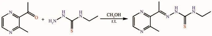
Scheme 1 Synthetic route of TSC ligand HL
5:Brown blocks.Yield:59%.Anal.Calcd.for C24H44N10O8S3Cu2(%):C,34.98;H,5.38;N,17.00.Found(%):C,34.79;H,5.22;N,17.21.FT-IR(KBr,cm-1):νN=C1 593,νN=C(pyrazine)1 536,νS=C795.
1.3 X-ray crystallography
The X-ray diffraction measurement for HL and complexes 1~5 was performed on a Bruker SMART APEXⅡCCD diffractometer equipped with a graphite monochromatized Mo Kα radiation(λ=0.071 073 nm)by using φ-ω scan mode.Semi-empirical absorption correction was applied to the intensity data using SADABS program[15].The structures were solved by direct methods and refined by full matrix least-square on F2using SHELXTL-97 program[16].All non-hydrogen atoms were refined anisotropically.All H atoms were positioned geometrically and refined using a riding model.Details of the crystal parameters,data collection and refinements are summarized in Table 1.
CCDC:1434105,HL;1434106,1;1434107,2;1434108,3;1434109,4;1434110,5.

Table 1 Crystal data and structure refinement for HL and complexes 1~5
2 Results and discussion
2.1 Crystal structures description
The diamond drawings of HL and complexes 1~5 are shown in Fig.1 and 2.Selected bond distances and angles are listed in Table 2.Hydrogen bond parameters are listed in Table 3.The bond length of C=S in free ligand is 0.168 0(2)nm,which is slightly shorter than that in complexes 2 and 3(0.168 5(5)~0.169 5(4)nm),showing that the TSC ligand is neutral in both complexes[17].On the contrary,the enolization of C=S in complexes 1,4 and 5 can be confirmed by the bond lengths of C-S in a range of 0.172 1(8)~0.175 6(4)nm,which are in excellent agreement with other previously known acylhydrazone complexes in the literature[17].In the crystal of HL,the molecules are linked into centrosymmetric dimers by pair of intermolecular N-H…O hydrogen bonds,forming a R22(10)ring motif.

Fig.1 ORTEP drawing of HL(a)and complexes 1~3(b~d)with 30%thermal ellipsoids
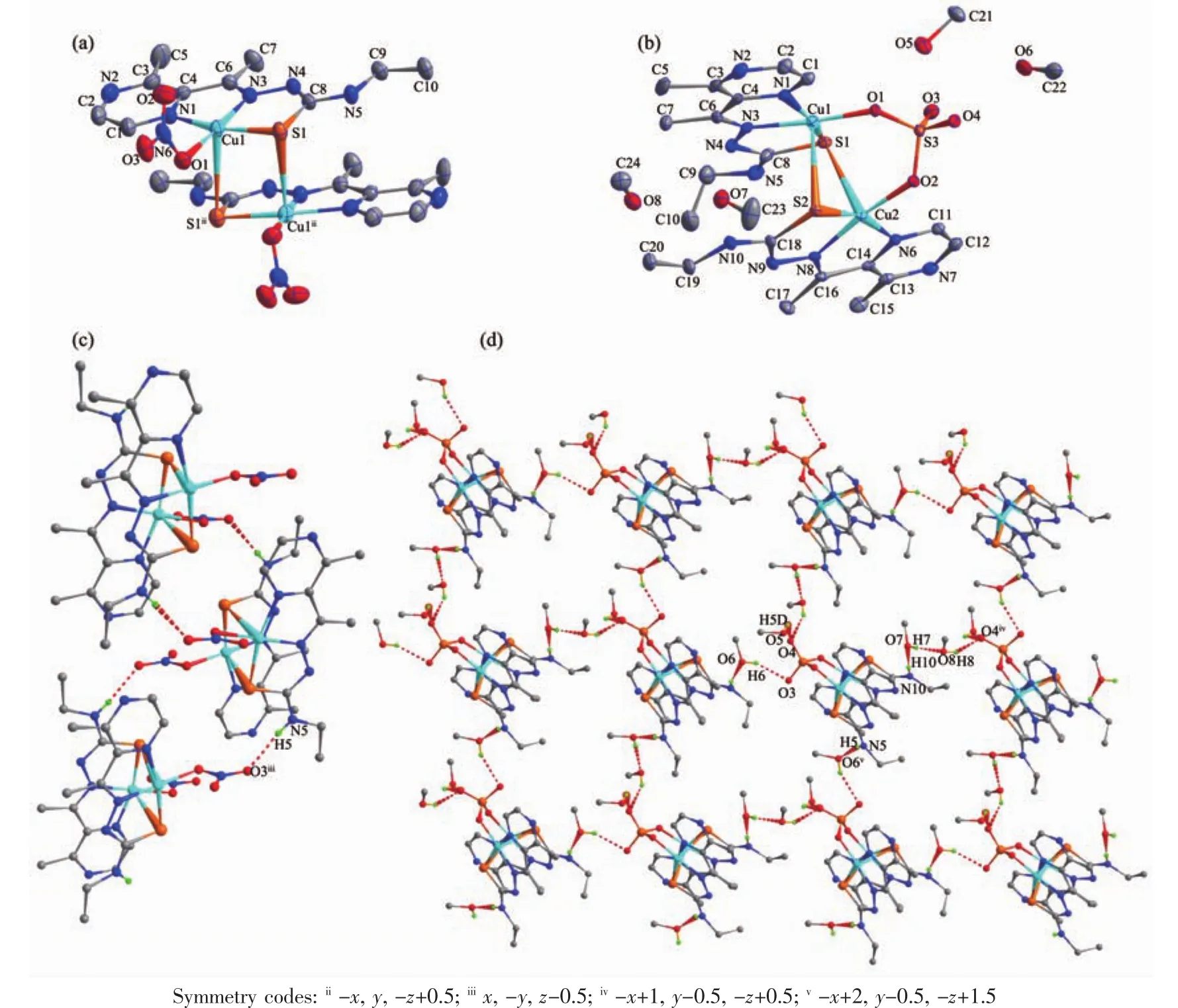
Fig.2 ORTEP drawing of complexes 4(a)and 5(b)with 30%thermal ellipsoids;Extended chain-like structure in the crystal of 4(c)and 2D supramolecular structure in the crystal of 5 formed by hydrogen bonds shown in dashed line(d)
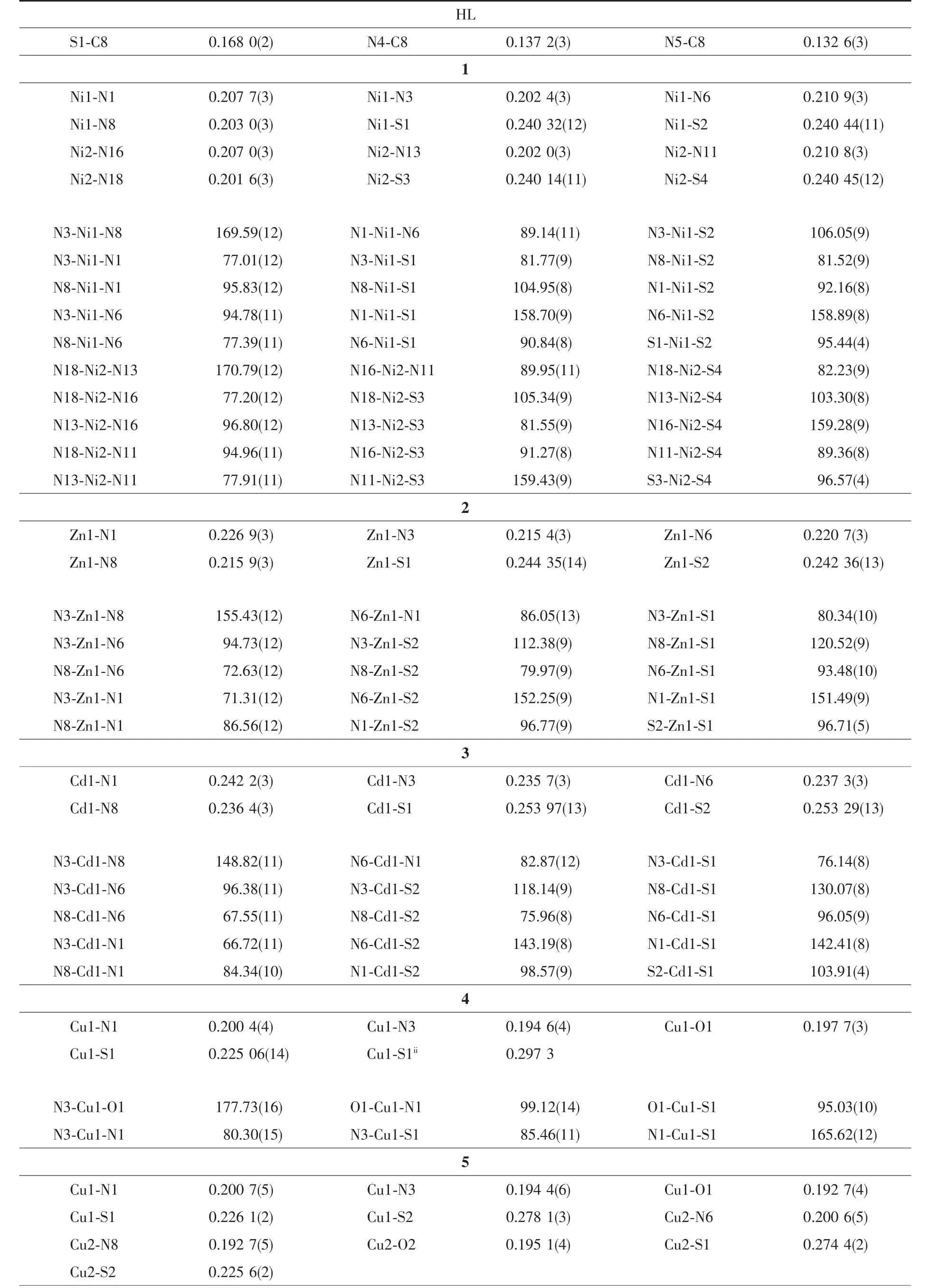
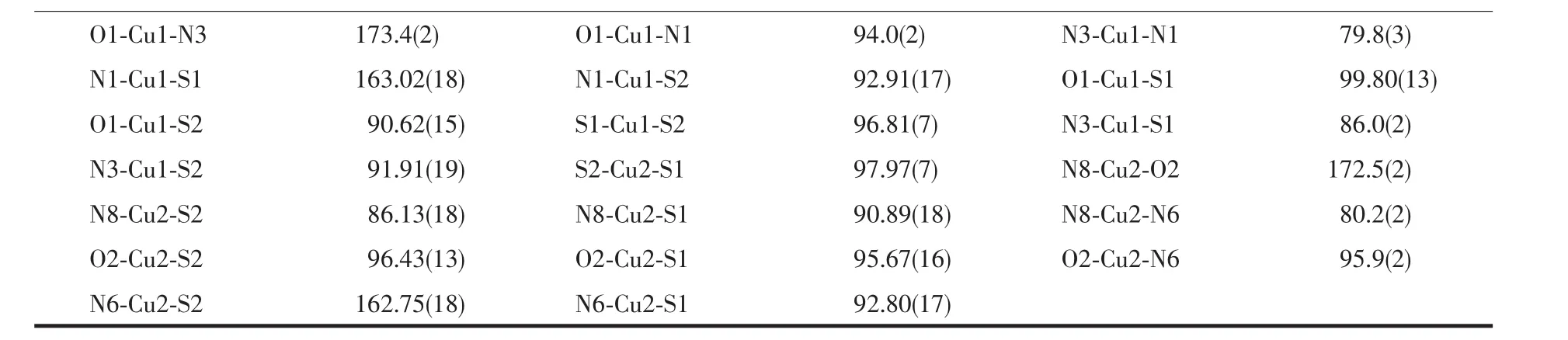
Table 2 Selected bond lengths(nm)and angles(°)for HL and complexes 1~5
Continued Table 1
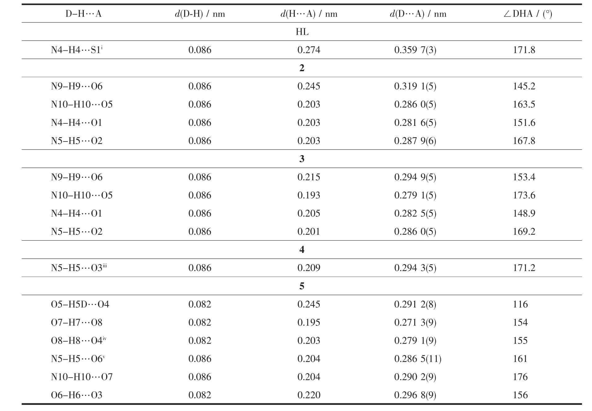
Table 3 Hydrogen bond parameters for HL and complexes 2~5
As shown in Fig.1a,1 is built of two independent neutral mononuclear molecules in the unit cell.Each NiⅡcenter is surrounded by two mono-anionic TSC ligands with N2S donor sets.The distances of Ni-N/S bonds are in a range of 0.201 6(3)~0.240 45(12)nm and are similar as those found in some reported complexes[18]. As expected, there is no classic hydrogen bonds in the crystal.
Complex 2 and 3 are isostructural,thus complex 2 is discussed for example.In complex 2(Fig.1b),the ZnⅡion is coordinated by two neutral TSC ligands with N2S donor sets,and thus possesses a distorted octahedron coordination geometry.There are two free nitrate anions in the outside of the complex for charge balance.In addition,intermolecular N-H…O hydrogen bonds between the complex cations and free nitrate anions could be observed in the crystals of 2 and 3.
Complex 4 crystallizes in a monoclinic space group C2/c,with one discrete dimeric CuⅡunit in the unit cell.As illustrated in Fig.2a,two Cu ions of the dimer are separated by 0.339 3 nm and doubly bridged by two S atoms of two TSC ligands to form a nearly planar four-membered Cu2S2core.Each of the CuⅡions is penta-coordinated by two S atoms from two different TSC ligands,two nitrogen atoms from one TSC ligand and one terminal bromide,giving a distorted square pyramid coordination geometry(τ=0.202).The distances of Cu-N/S bonds(0.194 6(4)~0.297 3 nm)are in good agreement with the corresponding distances reported in literature[18].In solid state,the discrete CuⅡdimers of 4 are further linked into a one-dimensional chain along c axis (Fig.2c)by intermolecular N-H…O hydrogen bonds.
X-ray analysis of 5 revealed that it also has a dimeric unit consisting of two CuⅡ,one SO42-ion and two TSC ligands,together with four crystal methanol molecules;it crystallizes in a monoclinic system with space group P21/c.As shown in Fig.2b,the coordination geometry around CuⅡin the dimeric complex is structurally similar to that of complex 4(τ=0.169 and 0.164 in the case of Cu1 and Cu2,respectively);the only difference is the presence of a μ2-SO42-at the outer axial sites on both CuⅡions.The four-membered Cu2S2core and the Cu-S distances(0.225 74(19)and 0.274 4(2)nm)are also similar to those found in complex 4.In the crystal,a large amount of intermolecular N-H…O and O-H…O hydrogen bonds link the molecules into a 2D supramolecular network(Fig.2d).
2.2 IR spectra
The most useful infrared spectral bands for determining the mode of coordination of ligands are the νN=C, νN=C(pyrazine),and νS=Cvibrations.The νN=Cband of the TSC ligand was found at 1 608 cm-1,while it shifted to 1 571~1 593 cm-1in complexes 1~5.The decrease in frequency of this band is an evidence for the coordination via the imine nitrogen atom[13,20].In the TSC ligand,a band at 1 553 cm-1is assigned to νN=C(pyrazine),whereas this band was found to shift to 1 533~1 536 cm-1in complexes 1~5,confirming that pyrazine nitrogen atom takes part in coordination[13].The νS=Cvibration at 813 cm-1in the TSC ligand shifted to lower frequency in the complexes,indicating the coordination of sulfur to the metal ions center[20].Additionally,bands at about 1 384 cm-1indicate that free nitrate groups exist in complexes 2 and 3.Two intense absorption bands in the spectra of complex 4 associated with the asymmetric stretching appeared at 1 474(ν1)and 1 343 cm-1(ν4),establishing that the coordinated NO3-groups are monodentate ligands[20].It is in accordance with the results of crystal structure analysis.
2.3 UV spectra
The UV spectra of the ligand HL,complexes 1~5 in CH3OH solution(c=10 μmol·L-1)were measured at room temperature(Fig.3).The spectrum of HL featured only one main band located around 308 nm (ε=31 806 L·mol-1·cm-1),which could be contributed to the characteristic π-π*transition of pyrazine[13].Similar bands were observed at 316 nm(ε=23 565 L·mol-1·cm-1),311 nm(ε=24 696 L·mol-1·cm-1),310 nm(ε=25 541 L·mol-1·cm-1),311 nm (ε=32 955 L·mol-1·cm-1)and 310 nm (ε=30 138 L·mol-1·cm-1)in complexes 1~5,respectively.However,there were new bands at 437 nm(ε=8 209 L·mol-1·cm-1),416 nm(ε=15 071 L·mol-1·cm-1),404 nm (ε=13 447 L·mol-1·cm-1),438 nm (ε=17 712 L·mol-1·cm-1)and 438 nm(ε =12 909 L·mol-1·cm-1)in the complexes 1 ~5,respectively,probably due to the ligand-to-metal charge transfer (LMCT)[21].All facts reveal the coordination of the ligand to the central metal ions.
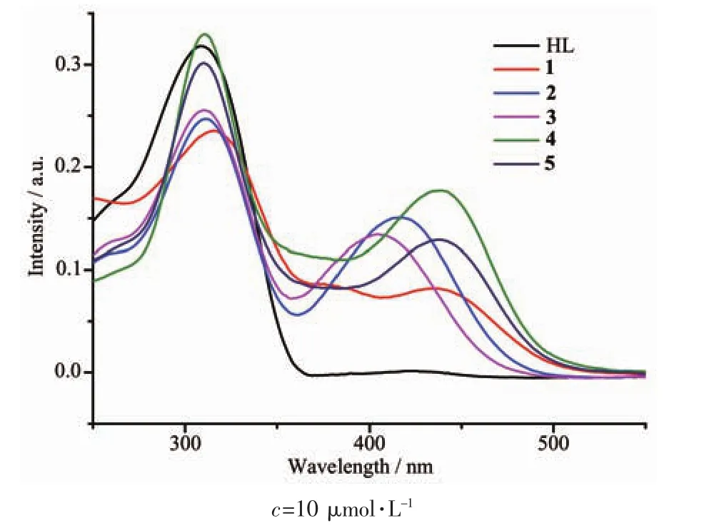
Fig.3 UV spectra of ligand HL,complexes 1~5 in CH3OH solution at room temperature
2.5 EB-DNA binding study by fluorescence spectrum
It is well known that EB can intercalate nonspecifically into DNA,which causes it to fluoresce strongly.Competitive binding of other drugs to DNA and EB will result in displacement of bound EB and a decrease in the fluorescence intensity[17].The effects of ligand HL and complexes 1~5 on the fluorescence spectra of EB-DNA system are presented in Fig.4.The fluorescence intensities of EB bound to ct-DNA at about 600 nm showed remarkable decreasing trends with the increasing concentration of tested compounds,indicating that some EB molecules are released into solution after the exchange with the compounds.The quenching of EB bound to DNA by the compounds is in agreement with the linear Stern-Volmer equation:I0/I=1+Ksqr[19],where I0and I represent the fluorescence intensities in the absence and presence of quencher,respectively;Ksqis the linear Stern-Volmer quenching constant,r is the ratio of the concentration of quencher and DNA.In the quenching plots of I0/I versus r,Ksqvalues are given by the slopes.The Ksqvalues are 1.069,0.824,0.752,0.947 and 1.107 for complexes 1~5,respectively,while that of the ligand HL is tested to be 0.587.The results indicate that the interactions of complexes 1~5 with DNA are stronger than that of ligand HL,which may be explained by the higher rigidity of the complexes to bind the base pairs along DNA[22].The NiⅡ complex 1,and CuⅡ complexes 4 and 5 displayed higher DNA interaction abilities than those of 2 and 3,indicating that the type of metal centers are responsible for the properties in some content.
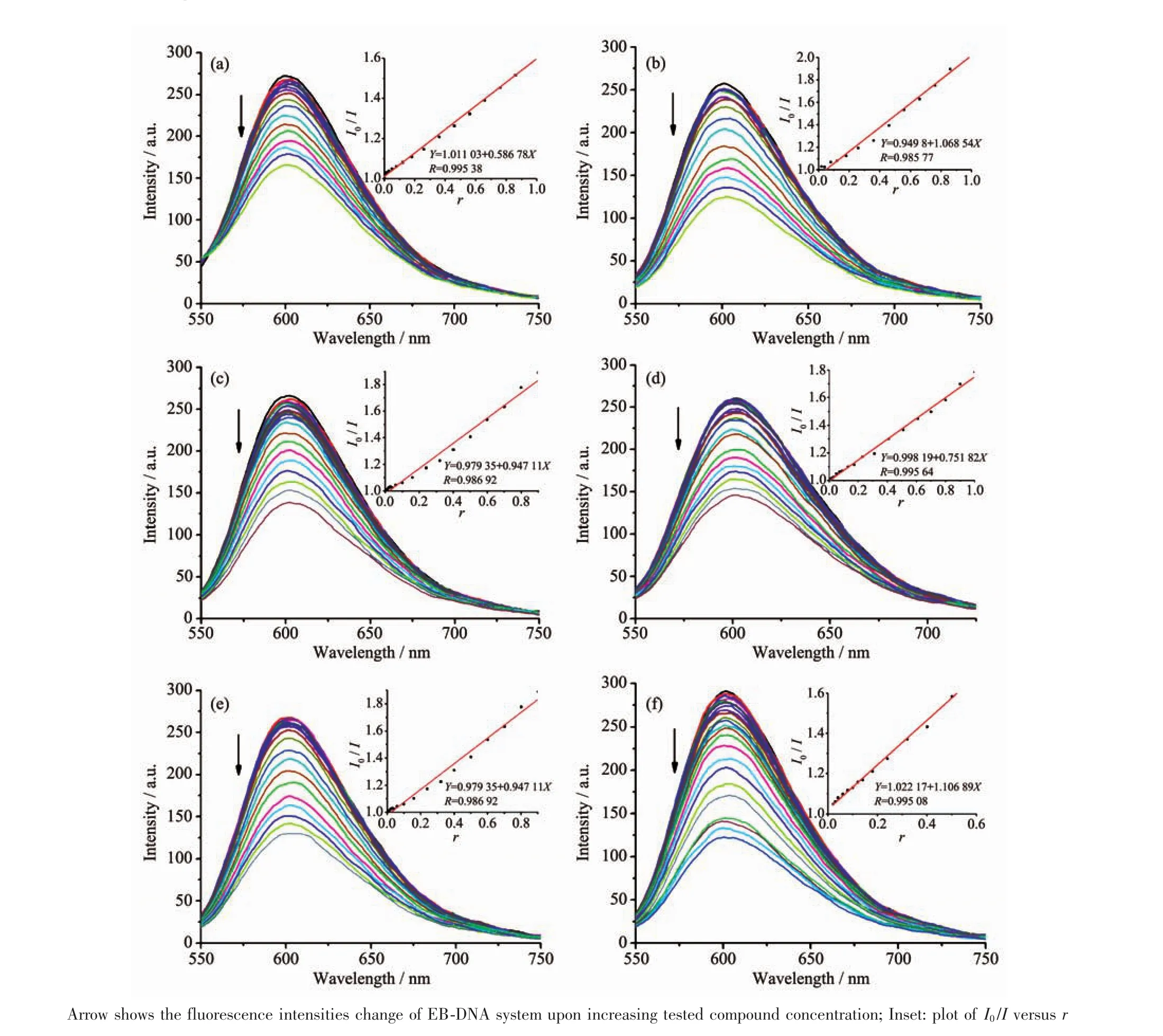
Fig.4 Emission spectra of EB-DNA system in the absence and presence of ligand HL(a)and complexes 1~5(b~f),respectively
3 Conclusions
Five transition metal complexes with a pyrazinecontaining thiosemicarbazone ligand were prepared and characterized by single-crystal X-ray crystallography.Moreover,the fluorescence spectra indicate that the interactions between the complexes and DNA are stronger than that of ligand HL,especially the NiⅡcomplex 1,and the dimeric CuⅡcomplexes 4 and 5,indicating thatthe type ofmetalcenters are responsible for the properties in some content.Further research is needed to better determine the relationship between structures and activities.
