程序性死亡配體-1與白細胞介素-10在喉鱗狀細胞癌中的表達及意義
2017-01-03 11:01:04葉惠平梁文卿李芊芊張安琪吳哲丹喻國凍
貴州醫藥 2016年10期
葉惠平 梁文卿 李芊芊 張安琪 吳哲丹 喻國凍
(貴州醫科大學附屬醫院耳鼻喉科,貴州 貴陽 550004)
?
程序性死亡配體-1與白細胞介素-10在喉鱗狀細胞癌中的表達及意義
葉惠平 梁文卿 李芊芊 張安琪 吳哲丹 喻國凍
(貴州醫科大學附屬醫院耳鼻喉科,貴州 貴陽 550004)
目的 通過檢測程序性死亡配體-1(PD-L1)和白細胞介素-10(IL-10)在喉鱗狀細胞癌中的表達情況,分析與喉鱗癌的腫瘤部位、病理分級、TNM分期、淋巴結轉移等之間的關系。方法應用免疫組化二步法檢測30例喉鱗狀細胞癌組織(觀察組)和15例癌旁正常組織(對照組)中PD-L1及IL-10的表達情況,分析二者與喉鱗狀細胞癌臨床病理參數的關系及二者表達的相關性。結果(1)PD-L1在喉鱗狀細胞癌組織(56.67%)中的表達明顯高于癌旁正常組織(13.33%);喉鱗狀細胞癌組織中PD-L1的陽性表達與病理分級、臨床分期(TNM分期)、T分期、腫瘤局部淋巴結轉移有關(P<0.05),與腫瘤分區、患者年齡無明顯相關性。(2)IL-10在喉鱗狀細胞癌組織(63.33%)中的表達明顯高于癌旁正常組織(20.00%);喉鱗狀細胞癌組織中IL-10的陽性表達與TNM分期、T分期、腫瘤局部淋巴結轉移有關(P<0.05),與病理分級、腫瘤分區、患者年齡無明顯相關性。(3)喉鱗狀細胞癌組織中PD-L1、IL-10的表達呈正相關(r=0.522,P<0.05)。結論(1)PD-L1與IL-10的高表達可能與喉鱗狀細胞癌的發病相關,兩者參與了腫瘤的發展及轉移;(2)免疫卡控點的調控和抑制性細胞因子與腫瘤的免疫逃逸密切相關,PD-L1與IL-10在喉鱗狀細胞癌中的表達呈正相關,兩者可能協同參與喉鱗狀細胞癌的免疫逃逸;(3)聯合檢測PD-L1與IL-10在喉鱗狀細胞癌組織中的表達對喉鱗癌的臨床評估有一定價值;為喉鱗癌的免疫治療提供理論支持。
程序性死亡配體-1; 白細胞介素-10; 喉鱗狀細胞癌
喉癌為耳鼻咽喉頭頸外科常見惡性腫瘤,發病率約占全身惡性腫瘤的1%~5%,近年來喉癌的發病有明顯升高及年輕化趨勢[1]。晚期喉癌由于浸潤和轉移,治療效果較差,其5年生存率約在60%以下[2]。近十年來免疫卡控點的研究,通過調控機體T細胞的免疫治療,使轉移性惡性腫瘤的治療獲得重大突破[3]。本研究以PD-L1及IL-10為對象,探討相關免疫指標在喉鱗狀細胞癌組織中的表達以及與喉鱗狀細胞癌患者臨床病理特征的相關性研究,為喉鱗狀細胞癌的免疫治療提供理論依據和思路。
1 資料與方法
1.1 組織標本 選取貴州醫科大學附屬醫院耳鼻咽喉頭頸外科2014年11月至2015年11月,經手術活檢后病理確診為喉鱗狀細胞癌標本,且術前均未行其它治療。男29例,女1例,年齡46~76歲,平均(62.17±11.18)歲。聲門上型9例,聲門型16例,聲門下型2例,貫聲門型3例。低分化1例,中分化17例,高分化12例。同時記錄患者TNM分期和淋巴結轉移情況。喉癌組織和癌旁正常黏膜置于10%中性緩沖福爾馬林固定,包埋及免疫組化檢測。
1.2 試劑與方法 兔抗人PD-L1單克隆抗體(ZA-0629)及免疫組化試劑盒(PV-6001)購自北京中杉金橋生物技術有限公司;兔抗人IL-10多克隆抗體(bS-0698)購自北京博奧森生物技術有限公司。本實驗采用非生物素二步法。
1.3 結果判斷 PD-L1和IL-10染色標準:本實驗觀察到PD-Ll及IL-10陽性染色主要定位于細胞膜及細胞質內。在400倍光學顯微鏡下隨機選擇5個無重復視野進行細胞計數,計算陽性細胞的比例,取其平均值。陽性細胞的百分比分級標準:陽性細胞≤10%為陰性(-),陽性細胞數≤30%為弱陽性(+),陽性細胞數≤60%為陽性(++),陽性細胞數>60%為強陽性(+++)。統計時將(+ ) ~(+++)均作為表達陽性。
1.4 統計學處理 采用SPSS 20.0統計軟件,組間差異比較用χ2檢驗,PD-L1與IL-10表達的相關性分析采用非參數Spearman秩相關分析。P<0.05為差異有統計學意義。
2 結 果
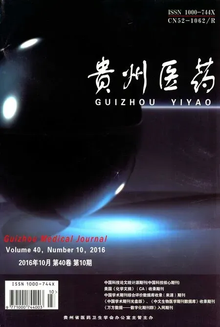
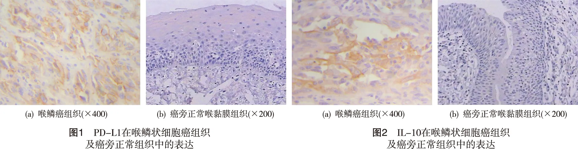
2.1 PD-L1、IL-10在喉鱗狀細胞癌組織和癌旁正常組織中表達 PD-L1陽性顯色表現呈棕黃色或棕褐色著色,喉鱗狀細胞癌組織中細胞膜和胞質中棕黃色顆粒大量聚集,腫瘤細胞著色明顯(圖1a),在部分正常組織中,亦發現PD-L1低表達,著色較淺,散在分布(圖1b);IL-10陽性顯色為棕黃色或棕褐色(圖2a),主要表達于細胞質及細胞膜,以細胞質表達為主(圖2b)。喉鱗狀細胞癌組織中PD-L1、IL-10的陽性表達率分別為56.67%(17/30)、63.33%(19/30),顯著高于PD-L1、IL-10在正常組織中的表達(P<0.05)。
2.2 PD-L1和IL-10在喉鱗狀細胞癌組織中的表達及其與臨床病理特征的關系 喉鱗狀細胞癌組織中PD-L1的陽性表達與病理分級、TNM分期、T分期、局部淋巴結轉移有關(P<0.05),PD-L1在中低分化喉鱗癌組和Ⅲ、Ⅳ期患者組及T3、T4期患者組的陽性表達率顯著高于臨床分期早期患者組;局部淋巴結轉移組PD-L1的陽性表達率87.50%明顯高于無淋巴結轉移組45.45%。喉鱗狀細胞癌組織中IL-10陽性表達與TNM分期、T分期、局部淋巴結轉移有關(P<0.05);IL-10在Ⅲ、Ⅳ期患者組、T3、T4期患者組、局部淋巴結轉移組中的陽性表達率分別為75.00%、 73.68%、 100.00%,但與病理分級、腫瘤分區、患者年齡無明顯相關性。
2.3 PD-L1和IL-10在喉鱗狀細胞癌組織中表達的相關性分析 PD-L1與IL-10在喉鱗狀細胞癌組織中的表達相關性檢驗采用Spearman秩相關分析,二者表達呈正相關,即隨著PD-L1表達增加,IL-10高表達所占的比例升高(rs=0.522,P<0.05)。
3 討 論
免疫卡控點的調控受到雙向機制調節[4],在T細胞與APC(抗原呈遞細胞)之間存在大量的配體受體結合的調控模式。這些細胞表面存在的受體和配體結合后,部分產生協調刺激信號,也有傳遞抑制信息。正常情況下,T細胞一旦出現活化,抑制信息往往會出現上調,避免對機體的過度攻擊。負調控途徑包括CTLA-4和CD80或CD60、PD-1和PDL-1、ICOS。其中程序性死亡受體-1,其最初是從凋亡的小鼠T細胞雜交瘤2B4.11克隆出來的[5]。PD-1可與其內源性配體PD-L1(B7-H1)或PD-L2(B7-DC)結合。研究[6-7]表明腫瘤微環境中呈現高度免疫抑制的狀態,且已在乳腺癌、前列腺癌、卵巢癌、黑色素瘤等多種癌癥的腫瘤浸潤性淋巴細胞(TILs)中發現了PD-1的高表達,且PD-1的高表達與腫瘤的分級、大小、淋巴結轉移、遠處轉移等有相關性,說明PD-1在腫瘤發生中具有重要作用,在多種腫瘤細胞中發現了PD-L1的高表達[8]。
本研究通過免疫組織化學染色顯示PD-L1在喉鱗狀細胞癌組織中主要表達于腫瘤細胞質中,PD-L1在喉鱗狀細胞癌組織中的表達明顯高于正常組織(P<0.05),我們推測在喉鱗狀細胞癌微環境中,PD-L1分子與受體PD-1分子介導的信號通路是介導喉鱗狀細胞癌免疫逃逸的重要信號分子。PD-L1在喉鱗狀細胞癌組織中異常高表達,與病理分級、TNM分期、T分期、腫瘤局部淋巴結轉移相關(P<0.05)。PD-L1在中低分化喉鱗癌中的陽性表達高于高分化癌組織,隨著更晚期的腫瘤分期和淋巴結轉移而增加,說明PD-L1在喉鱗癌細胞中的表達變化是構成喉鱗癌細胞微環境的非常重要的因素,伴隨著喉鱗狀細胞癌發病、惡性發展及轉移的整個病程。
IL-10是腫瘤發生發展過程中一種重要的免疫抑制因子。腫瘤細胞自身也可分泌大量IL-10。IL-10主要通過以下幾方面參與抑制機體抗腫瘤免疫反應:一方面,IL-10抑制Th1分化和細胞因子的合成。機體的抗腫瘤免疫主要通過Th1亞群介導的細胞免疫來完成,IL-10通過抑制抗原提呈細胞表面MHC-Ⅱ類分子和協同刺激分子CD80和CD86的表達,從而抑制抗原提呈給T細胞的過程,間接實現對Th1細胞的抑制;同時,IL-10抑制樹突細胞產生Th1細胞分化所必需的細胞因子IL-12來抑制Th1的分化。另一方面,IL-10可以抑制CTL的增殖和細胞毒作用[9]。活化的CTL的主要作用是作為免疫效應分子誘導腫瘤細胞的凋亡,IL-10通過抑制Th1細胞和單核/巨噬細胞產生的細胞因子,從而抑制CTL的活化和分化;IL-10還可以抑制腫瘤細胞表面MHC-Ⅰ類抗原的表達,從而影響CTL對腫瘤抗原的識別,使腫瘤細胞逃避CTL的殺傷作用[10]。
本研究通過免疫組織化學方法檢測到IL-10在喉鱗狀細胞癌組織中的表達明顯高于其在正常組織中的表達,且IL-10的陽性表達與TNM分期、T分期、腫瘤局部淋巴結轉移有關(P<0.05),在更晚期腫瘤分期及有淋巴結浸潤的喉鱗癌組織中更易檢測到,這表明IL-10在喉鱗狀細胞癌發生、發展及轉移中起作用。Dulos等[11]發現PD-1能夠抑制CD4+T細胞向Th1亞群分化,而促進其向Th2亞群分化。文獻[12-13]發現,PD-L1能夠增強Treg的功能,并且促進Treg產生IL-10。結合本研究結果,喉鱗狀細胞癌組織中PD-L1與IL-10的表達呈正相關(P<0.05),兩者可能共同參與喉鱗狀細胞癌免疫逃避過程。
我們研究發現PD-L1與IL-10的高表達可能與喉鱗狀細胞癌的發病相關,促進了腫瘤的惡性發展及頸部淋巴結轉移。免疫卡控點的調控和免疫抑制因子與腫瘤的免疫逃逸密切相關,PD-L1與IL-10在喉鱗狀細胞癌中的表達呈正相關,可能協同參與喉鱗狀細胞癌的免疫逃逸,為喉癌的免疫治療提供理論依據,調控喉鱗癌組織中的上述靶點,有望成為控制喉鱗狀細胞癌惡性發展、淋巴結轉移及復發的新途徑。
[1] Hoffman H T,Karnell L H,Funk G F,et al.The National Cancer Data Base report on cancer of the head and neck[J].Archives of otolaryngology-head & neck surgery,1998,124(9):951-962.
[2] Spector G J,Sessions D G,Jason Lenox M S,et al.Management of stage IV glottic carcinoma:therapeutic outcomes[J].Laryngoscope,2004,114(8):1438-1446.
[3] Callahan MK,Postow MA,Wolchok JD.Immunomodulatory therapy for melanoma:ipilimumab and beyond[J].Clin Dermatol,2013,31:191-199.
[4] Zou W C,Inhibitory L.B7-family molecules in the tumour microenvironment[J].Nature Rev Immunol,2008,8:447-467.
[5] Ishida Y,Agata Y,Shibahara K,et al.Induced expression of PD-1,a novel member of the immunoglobulin gene superfamily,upon programmed cell death[J].The EMBO Journal,1992,11(11):3887-3895.
[6] Lauerova L,Dusek L,Simickova M I,et al.Malignant melanoma associates with Th1/Th2 imbalance that coincides with disease progression and immunotherapy response[J].Neoplasma,2002,49(3):159-166.
[7] Mirjana U,Joerg W,Beatrix M,et al.HLA-G protein up-regulation in primary cutaneous lymphomas is associated with interleukin-10 expression in large cell T-cell lymphomas and indolent B-cell lymphomas.[J].Blood,2002,2(3):179-197.
[8] Sheu B C,Lin R H,Lien H C,et al.Predominant Th2/Tc2 polarity of tumor-infiltrating lymphocytes in human cervical cancer.[J].Journal of Immunology,2001,167(5):2972-2978.
[9] Sharma S,Stolina M,Lin Y,et al.T cell-derived IL-10 promotes lung cancer growth by suppressing both T cell and APC function.[J].Journal of Immunology,1999,163(9):5020-5028.
[10] Petersson M,Charo J,Salazar-Onfray F,et al.Constitutive IL-10 production accounts for the high NK sensitivity,low MHC class I expression,and poor transporter associated with antigen processing (TAP)-1/2 function in the prototype NK target YAC-1[J].Journal of Immunology,1998,161(5):2099-2105.
[11] Dulos J,Carven G J,VanBoxtel S J,et al.PD-1 blockade augments Th1 and Th17 and suppresses Th2 responses in peripheral blood from patients with prostate and advanced melanoma cancer[J].Journal of Immunotherapy,2012,35(2):169-178.
[12] Qing D,Liming L,Xiaorong Z,et al.Human PD-L1-overexpressing porcine vascular endothelial cells induce functionally suppressive human CD4+CD25hiFoxp3+Treg cells[J].Journal of Leukocyte Biology,2011,90(1):77-86.
[13] Seung-Pil S,Hye-Hyun S,Jae-Hun S,et al.Adenovirus expressing both thymidine kinase and soluble PD1 enhances antitumor immunity by strengthening CD8 T-cell response[J].Molecular Therapy the Journal of the American Society of Gene Therapy,2013,21(3):688-695.
Expression and Role of Programmed death-1 ligand and Interleukin-10 in Laryngeal Squamous Cell Carcinomas
YeHuiping,LiangWenqing,LiQianqian,ZhangAnqi,WuZhedan,Yuguodong.
DepartmentofEar-Nose-Throat,GuizhouMedicalUniversity,Guiyang550004,Guizhou,China.
Objective To identify the relationship between the expression of programmed death-1 ligand (PD-L1) and interleukin-10 (IL-10) and the tumorous division, clinical stage, pathological grade and lymph node metastasis of LSCC.Methods The expression of PD-L1 and IL-10 were detected by two-step immunohistochemistry in thirty cases of LSCC tissues (experiment group) and fifteen cases of para-carcinoma tissues (control group), the correlation between them and their correlations with the clinicopathological parameters were analyzed. Results The levels of PD-L1 positive expression in LSCC tissue (56.67%) were significantly higher than that of histologically normal tissue (13.33%). In the LSCC tissues, the expression of PD-L1 was significantly associated with the pathological grade, T stage, TNM stage of LSCC and lymph node metastasis (P<0.05), but no association was observed with tumor sites and age of patients. The levels of IL-10 expression in LSCC tissue (63.33%) were significantly higher than that of histologically normal tissue (20.00%). In the LSCC tissues, the expression of PD-L1 was significantly associated with the T stage, TNM stage of LSCC and lymph node metastasis (P<0.05), but no association was observed with pathological grade, tumor sites and age of patients. The expression of PD-L1 pro-correlated with IL-10 in the LSCC tissues (r=0.522,P<0.05). Conclusion The high expression of PD-L1 and IL-10 may be closely related to the occurrence, development and metastasis of LSCC. The expression of PD-L1 pro-correlated with IL-10 in the LSCC tissues, they may be interactive with each other in the immune escape of LSCC together. Inhibition of their expression in LSCC might be a new treatment to LSCC.
Programmed death-1 ligand; Interleukin-10; Laryngeal squamous cell carcinomas
貴州省科技廳聯合基金項目資助(LG-2012-068);貴州醫科大學附屬醫院博士啟動基金資助
R739.65
A
1000-744X(2016)10-1019-03
2016-06-23)