山羊顳下頜關節盤膠原構筑和生物力學性能研究
2016-07-22 10:40:47李燕梅包廣潔2鐘妮呂瑋張文霞王蘭蘭康宏
華西口腔醫學雜志 2016年1期
李燕梅包廣潔,2鐘妮呂瑋張文霞王蘭蘭康宏
1.蘭州大學口腔醫學研究所,蘭州 730000;2.西北民族大學口腔醫學國家民委重點實驗室,蘭州 730030;3.蘭州大學生命科學學院,蘭州 730000
?
山羊顳下頜關節盤膠原構筑和生物力學性能研究
李燕梅1包廣潔1,2鐘妮1呂瑋1張文霞1王蘭蘭3康宏1
1.蘭州大學口腔醫學研究所,蘭州 730000;2.西北民族大學口腔醫學國家民委重點實驗室,蘭州 730030;3.蘭州大學生命科學學院,蘭州 730000
[摘要]目的 為工程化顳下頜關節盤組織建立客觀的生物力學標準。方法 從4只健康1月齡山羊頭中完整分離8個顳下頜關節盤,用直徑3 mm的沖子在前、中、后帶取圓柱狀試樣,預處理壓縮速度為0.025 mm·s-1,結束條件為最大載荷小于4.9 N,壓縮位移1.5 mm,循環3次,在預處理基礎上完成壓縮實驗,獲取各區帶的應力應變關系圖和彈性模量值。掃描電子顯微鏡和偏振光顯微鏡觀察各區帶的超微結構和膠原纖維走向。結果 1)關節盤組織的生理應變范圍在10%以內,關節盤組織的擬線性本構方程可以用多項式y=ax+bx2+cx3進行擬合。2)關節盤中間帶的彈性模量值最大,其與前帶和后帶均有統計學差異(P<0.05),前帶與后帶之間無統計學差異(P=0.361)。3)顳下頜關節盤內部呈現環形膠原網狀結構,中間帶膠原排列致密,呈前后向排列;前帶和后帶主要呈內外走向,伴有前后方向的纖維。結論 環形膠原網狀結構是山羊顳下頜關節盤抗力性能的基礎,中間帶抗壓性能高于前帶和后帶。
[關鍵詞]顳下頜關節盤; 擬線性本構方程; 壓縮實驗; 彈性模量; 掃描電子顯微鏡; 偏振光顯微鏡
顳下頜關節盤(temporomandibular joint disc,TMD)是下頜髁突和顴骨關節窩之間的纖維軟骨樣組織,主要由成纖維樣細胞和軟骨樣細胞構成。國外研究[1]報道,50%~75%的人至少有過單側顳下頜關節疼痛的經歷,其中33%的人至少有一次持續性關節疼痛癥狀。我國顳下頜關節盤的既往功能紊亂患病率為18.3%,但臨床檢查功能紊亂患病率為54.2%,女性高于男性[2-3]。學者[4-5]認為顳下頜關節病可能是由精神因素、因素、其他因素(如行為習慣、創傷、自然環境、代謝、免疫、關節形態發育異常等)綜合導致,其中大約70%的關節疾病與關節盤的病變有關,多以關節盤變薄、透明樣變、穿孔為主要特征。正常情況下,顳下頜關節盤具有良好的抗壓特性,但在受到不正常的壓力或穿孔后,一般不能自我修復。近年來,組織工程化顳下頜關節盤的研究為臨床治療關節盤疾病提供了一條新途徑,但是這一領域的研究存在的主要問題有:沒有理想的細胞源和合適的動物模型、支架的生物學功能匹配性差、工程化組織的強度和功能與正常組織有較大差異等,制約了這項工作的開展[6]。
研究[7-8]表明,山羊的顳下頜關節盤在形態、結構和功能方面較其他動物更接近于人。筆者在前期的實驗中對山羊關節盤的形態學、組織學以及免疫組織化學進行了初步分析[9],證明用山羊作為組織工程化顳下頜關節盤研究的動物模型是可行的[10]。對山羊關節盤的生物力學性能進行表征,將為工程化顳下頜關節盤組織建立客觀的評價標準,為功能性組織工程的深入研究提供依據。
1 材料和方法
1.1 主要儀器
Instron 5960系列雙立柱臺式微力試驗機(Instron公司,美國),是一款電子萬能微力試驗機,可用于各種組織原材料、復合材料、塑料、橡膠材料和金屬等的壓縮、拉伸以及剪切力學性能的測試,與Bluehill 3軟件配套使用。
1.2 標本收集
從當地屠宰場購買新鮮屠宰的健康1月齡山羊頭4個,清洗干凈后在75%乙醇中浸泡半小時,然后在超凈工作臺內完整切取雙側顳下頜關節盤,將周圍關節囊組織修剪干凈后,用生理鹽水徹底沖洗備用。
1.3 生物力學測試
1.3.1 試樣制備 根據文獻[11]中關節盤的五分區方法,將關節盤分為前、中、后、內、外帶,每個區帶大小均為關節盤的五分之一,并在前、中、后帶中央按冠狀方向用取樣工具沖子(直徑3 mm)制作圓柱狀壓縮試樣,每帶取4個試樣放入4 ℃冰箱中備用。
1.3.2 預處理 實驗前將試樣從4 ℃冰箱中取出,用數顯卡尺測出每個試樣的厚度值,在微力試驗機上輸入基本參數(樣品厚度和直徑)。打開Bluehill 3軟件,設置預處理壓縮速度為0.025 mm·s-1,結束條件為最大載荷小于4.9 N,壓縮位移小于1.5 mm[11-12]。壓縮過程中,將試樣置于一次性培養皿中,并滴注少量PBS液保持試樣濕潤,手動調節壓頭,壓頭與試樣無限接近時,開始預壓縮處理,循環3次即可。
1.3.3 壓縮實驗 在預處理的基礎上設置實驗方法和實驗參數,實驗載荷從零緩慢增加至其最大值(小于4.9 N,傳感器精度為5 N),利用Bluehill 3軟件計算出試樣的彈性模量值、壓縮應變在10%和20%時的壓縮應力、載荷、時間、位移等參數[12-14],獲取各區帶的應力應變的關系圖。
1.4 掃描電子顯微鏡和偏振光顯微鏡觀察
關節盤組織按不同分區制備成約2 mm大小的樣品,每個分區各4個樣品。在4%戊二醛中浸泡24 h,常規梯度乙醇脫水,冷凍干燥機中干燥8 h,真空噴金,在掃描電子顯微鏡(S-3400N型,Hitachi公司,日本)下觀察關節盤不同分區的表面和斷面超微結構。根據試樣分區,利用冰凍切片機(CM1100型,LEICA公司,德國)將關節盤試樣按水平面和冠狀面切成20 μm厚的切片,在偏振光顯微鏡(BX51型,Olympus公司,日本)下觀察關節盤內部不同區帶的膠原纖維走向。
1.5 統計學處理
采用SPSS 19.0軟件進行統計分析。單因素方差分析(ANOVA)和LSD-t檢驗比較關節盤前、中、后帶的彈性模量的差異,選取α值為0.05。
2 結果
2.1 關節盤不同區間壓縮應力與應變的關系
從關節盤前、中、后帶的應力應變曲線圖(圖1)可見,各帶的曲線圖走形基本一致,坡腳區(toeregion)出現在壓縮應變的10%以內,即關節盤組織的生理應變范圍在10%以內。壓縮應變達到10%以后,曲線變陡直,應力隨應變明顯增加。各區帶的坡腳區略有不同,中間帶的坡腳區相對較小。
2.2 關節盤不同區間壓縮應力與應變的理論分析
根據擬線性黏彈性理論,關節盤各區帶的應力應變值可以用多項式y=ax+bx2+cx3作為擬線性本構方程,得到擬合后的實驗曲線。此本構方程可以為顳下頜關節盤組織工程提供原始力學性能參考。
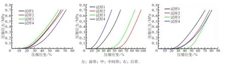
圖 1 關節盤各帶的應力-應變曲線圖Fig 1 Stress-strain curve of articular disc of each band
2.3 關節盤不同區間彈性模量的比較
山羊關節盤前、中、后帶的彈性模量分別為(2.21±0.14)、(2.47±0.11)、(2.25±0.19) MPa。統計分析表明,關節盤中間帶的彈性模量值最大,其與前帶和后帶均具有統計學差異(P=0.002,P= 0.018),前帶與后帶間無統計學差異(P=0.361)。
2.4 掃描電子顯微鏡和偏振光顯微鏡分析
關節盤各帶的掃描電子顯微鏡和偏振光顯微鏡分析見圖2和圖3。
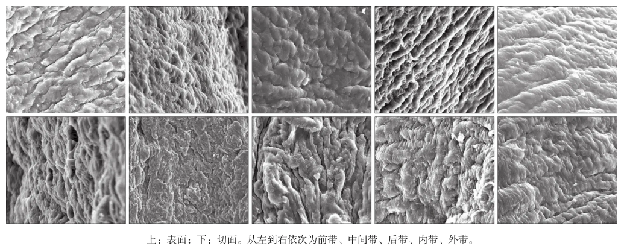
圖 2 關節盤各帶的表面和切面 掃描電子顯微鏡 × 1 000Fig 2 Articular disc surface and cut surface of each band scanning electron microscope × 1 000
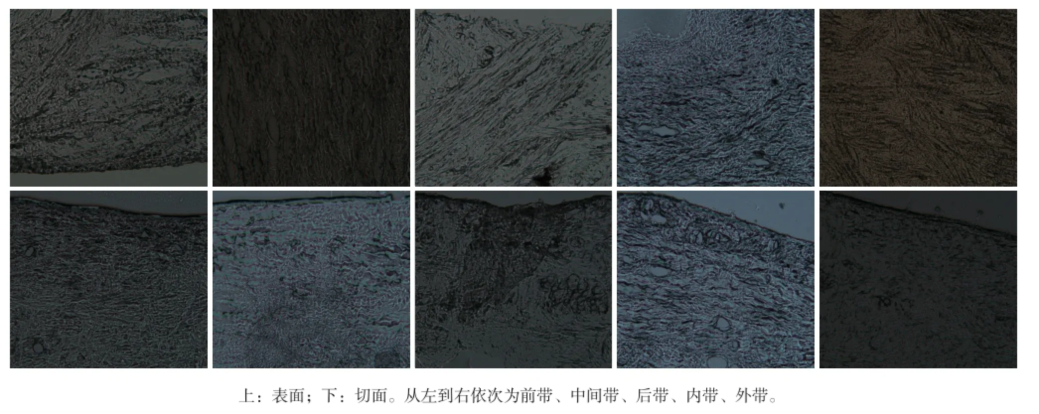
圖 3 關節盤各帶的表面和切面 偏振光顯微鏡 × 200Fig 3 Articular disc surface and cut surface of each band polarized light microscope × 200
掃描電子顯微鏡(圖2)可見,關節盤表面均勻覆蓋著凝膠樣物質,纖維粗細從幾微米到幾十微米不等。水平面掃描顯示,前帶和后帶的纖維大致平行排列呈波紋或溝紋狀結構,而中間帶的膠原纖維束則排列致密,呈條索狀,關節盤內外側的膠原纖維束呈網紋狀排列,外側較內側粗。切面掃描顯示,前、后、內、外帶纖維方向不完全單一而呈現參差不齊的纖維斷面,中間帶則呈現出魚鱗狀的纖維束斷面。偏振光顯微鏡圖(圖3)可見,在山羊顳下頜關節盤內部,其周圍區域纖維呈環形包繞而在中間帶區域呈前后向排列,前帶和后帶區域的纖維主要呈內外走向并伴有前后方向的分支纖維,內側和外側的纖維以前后走向為主,夾雜有上下和內外向的分支纖維。綜合分析,顳下頜關節盤內部呈現環形膠原網狀結構。
3 討論
彈性模量是表征在彈性限度內物質材料抗壓或抗拉的物理量,其大小標志了材料的剛性,模量越大,越不容易發生形變[15]。本實驗顯示,山羊關節盤中間帶的彈性模量大于前帶和后帶,說明中間帶的剛性最大,抵抗壓縮或外界沖擊力的性能也最好。掃描電子顯微鏡和偏振光顯微鏡觀察顯示,中間帶以致密的前后向膠原纖維為主,與其他區域的三維網狀結構有所不同。
根據關節軟骨二相理論,顳下頜關節盤是由黏彈性固體基質(膠原和蛋白多糖)和自由流動的間隙液組成的二相混合物,在生物力學性能方面具有彈性和黏彈性。本研究結果表明,關節盤在承受較小的應力應變范圍(10%)內,發生的彈性形變可以恢復到原來的狀態,這一區域即為組織的生理應變區,在應力應變圖上顯示為坡腳區。但當超過應變的10%,組織的剛性隨著應變的變化迅速增加,反映了組織通過對剛度的調節來適應外界條件的變化。本實驗結合擬線性黏彈性理論,采用多項式對不同區帶的應力應變曲線進行理論擬合,獲得了多項式方程y=ax+bx2+cx3,這為今后組織工程化顳下頜關節盤的研究提供了方法和實驗依據。
本實驗按不同分區取樣進行了掃描電子顯微鏡和偏振光顯微鏡的觀察,結果發現,山羊顳下頜關節盤中間帶纖維呈前后走向的致密板層狀結構,前帶和后帶的纖維一般為內外向并穿插有前后向和上下向的細小纖維束。組織的抗壓縮性能與其內的組織液含量有很大關系,山羊關節盤中間帶的板層樣結構對外界的沖擊壓力有一定的對抗和緩沖作用,這與其他學者[16]對豬顳下頜關節盤內的生物力學功能的研究結果接近。此外,關節盤的抗壓縮性能除了與其內的液體含量、膠原纖維分布和走向有關外,可能還與關節盤不同區帶間富含的蛋白多糖和糖胺多糖的含量有關。對豬關節盤不同區帶的定量生化研究顯示,中間帶含有最多的膠原蛋白和糖胺多糖[16],這可能也是關節盤中間帶彈性模量高的原因之一。
[參考文獻]
[1]Reneker J, Paz J, Petrosino C, et al. Diagnostic accuracy of clinical tests and signs of temporomandibular joint disorders: a systematic review of the literature[J]. J Orthop Sports Phys Ther, 2011, 41(6):408-416.
[2]劉洪臣. 我國顳下頜關節病的研究與臨床進展[J]. 中華口腔醫學雜志, 2014, 49(7):385-389. Liu HC. Research and clinical progression of temporomandibular joint disease in China[J]. Chin J Stomatol, 2014, 49 (7):385-389.
[3]傅開元. 診治顳下頜關節紊亂病必須明確的問題[J]. 中華口腔醫學雜志, 2014, 49(10):581-584. Fu KY. The problem of diagnosis and treatment of temporomandibular joint disorder[J]. Chin J Stomatol, 2014, 49 (10):581-584.
[4]Murphy MK, MacBarb RF, Wong ME, et al. Temporomandibular disorders: a review of etiology, clinical management,and tissue engineering strategies[J]. Int J Oral Maxillofac Implants, 2013, 28(6):e393-e414.
[5]Iodice G, Danzi G, Cimino R, et al. Association between posterior crossbite, masticatory muscle pain, and disc displacement: a systematic review[J]. Eur J Orthod, 2013, 35 (6):737-744.
[6]Shu W, Liu L, Bao G, et al. Tissue engineering of the temporomandibular joint disc: current status and future trends [J]. Int J Artif Organs, 2015, 38(2):55-68.
[7]Cheung LK, Shi XJ, Zheng LW. Surgical induction of temporomandibular joint ankylosis: an animal model[J]. J Oral Maxillofac Surg, 2007, 65(5):993-1004.
[8]Wang YL, Li XJ, Qin RF, et al. Matrix metalloproteinase and its inhibitor in temporomandibular joint osteoarthrosis after indirect trauma in young goats[J]. Br J Oral Maxillofac Surg, 2008, 46(3):192-197.
[9]舒維娜, 康宏, 張衛平, 等. 山羊顳下頜關節盤細胞體外培養研究[J]. 實用口腔醫學雜志, 2010, 26(2):165-168. Shu WN, Kang H, Zhang WP, et al. Isolation and cultiva-tion of goat temporomandibular joint disc cells[J]. J Pract Stomatol, 2010, 26(2):165-168.
[10]康宏, 李振強, 閉艷妲. 自組裝山羊顳下頜關節盤組織工程纖維軟骨模型的構建[J]. 華西口腔醫學雜志, 2011, 29 (3):314-317. Kang H, Li ZQ, Bi YD. Self-assembly tissue engineering fibrocartilage model of goat temporomandibular joint disc [J]. West China J Stomatol, 2011, 29(3):314-317.
[11]舒維娜, 康宏, 張衛平, 等. 山羊顳下頜關節盤細胞類型及分布表征在組織構建中的設計意義[J]. 中國組織工程研究與臨床康復, 2009, 13(46):9022-9026. Shu WN, Kang H, Zhang WP, et al. Type and distribution of cells in goat temporomandibular joint discs and its significance in tissue engineering design[J]. J Clin Rehabil Tissue Eng Res, 2009, 13(46):9022-9026.
[12]Hadidi P, Athanasiou KA. Enhancing the mechanical properties of engineered tissue through matrix remodeling via the signaling phospholipid lysophosphatidic acid[J]. Biochem Biophys Res Commun, 2013, 433(1):133-138.
[13]Hadidi P, Yeh TC, Hu JC, et al. Critical seeding density improves the properties and translatability of self-assembling anatomically shaped knee menisci[J]. Acta Biomater, 2015,11:173-182.
[14]Juran CM, Dolwick MF, McFetridge PS. Shear mechanics of the TMJ disc: relationship to common clinical observations[J]. J Dent Res, 2013, 92(2):193-198.
[15]MacBarb RF, Chen AL, Hu JC, et al. Engineering functional anisotropy in fibrocartilage neotissues[J]. Biomaterials, 2013,34(38):9980-9989.
[16]Detamore MS, Orfanos JG, Almarza AJ, et al. Quantitative analysis and comparative regional investigation of the extracellular matrix of the porcine temporomandibular joint disc[J]. Matrix Biol, 2005, 24(1):45-57.
(本文編輯 李彩)
[中圖分類號]R 322.4+1
[文獻標志碼]A [doi] 10.7518/hxkq.2016.01.015
[收稿日期]2015-07-24; [修回日期] 2015-10-27
[基金項目]國家自然科學基金(81160139)
[作者簡介]李燕梅,碩士,E-mail:ymli13@lzu.edu.cn
[通信作者]康宏,教授,博士,E-mail:Kanghong@lzu.edu.cn
Collagen structure and biomechanical properties of the goat temporomandibular joint disc
Li Yanmei1, Bao Guangjie1,2,Zhong Ni1, Lü Wei1, Zhang Wenxia1, Wang Lanlan3, Kang Hong1.
(1. Institute of Stomatology, Lanzhou University, Lanzhou 730000, China; 2. Key Lab of Stomatology of State Ethnic Affairs Commission, Northwest University for Nationalities, Lanzhou 730030, China; 3. School of Life Science, Lanzhou University, Lanzhou730000, China)
Supported by: The National Natural Science Foundation of China (81160139). Correspondence: Kang Hong, E-mail: Kanghong@lzu.edu.cn.
[Abstract]Objective To establish objectively biomechanical criteria for temporomandibular joint disc tissue engineerings. Methods Eight temporomandibular joint discs from a four-month-old goat were separated completely. A cylindrical sample with diameter of 3 mm in the anterior, intermediate, and posterior bands was obtained, and the samples underwent pre-compression test with three cycles under a speed of 0.025 mm·s-1. With the end condition for the maximum load of less than 4.9 N and 1.5 mm compression displacement, the formal compression test was performed with the same speed. Moreover, this test obtained the stress-strain relationship and elastic modulus of each disc band. Ultrastructure and collagen fiber orientation of the district zone were observed by scanning electron microscope and polarized light microscope. Results 1) The physiological strain range of an articular disc was within 10%, and the quasilinear constitutive equation of articular disc tissues can be fitted with the polynomial function: y=ax+bx2+cx3. 2) The elastic modulus of the intermediate disc zone was the largest (P<0.05). No significant difference existed between the anterior and posterior bands (P=0.361). 3) Scanning electron microscope and polarized light microscope showed an annular disc collagen network structure, which was the internal part of the goat temporomandibular joint disc. The collagen arrangement of intermediate bands was tensely arranged anterior-posteriorly. The collagen of anterior and posterior bands went through mediolaterally with intersection of anterior-posterior branch fibersd. Conclusion Annular collagen network structure is the basis for goat temporomandibular joint disc compression resistance properties. The intermediate band demonstrated higher compression resistance performance than the anterior and posterior bands.
[Key words]temporomandibular joint disc; quasi linear constitution equation; compressive testing; elastic modulus; scanning electron microscope; polarized light microscope