Value of diffusion-weighted MR imaging and dynamic-contrast enhanced MRI in the diagnosis of breast cancer
2012-10-23 02:05:32MangrooAteelesh艾提拉什LizhiYu李志宇ZhiYanHe之彥何WangPeijun王培軍
外科研究與新技術 2012年1期
Mangroo Ateelesh(艾提拉什),Lizhi Yu(李志宇),Zhi-Yan He(之彥何),Wang Peijun(王培軍)
Department of Radiology,Tongji Hospital of Tongji University,Shanghai 200065,China
1 Introduction
1.1 MRI is a well-recognized tool for detection,diagnosis and staging of breast cancer[1-3].MRI has been shown to minimize costs in some clinical settings by limiting or eliminating the need for further expensive or more invasive diagnostic or surgical procedures[4,5].
1.2 Diffusion imaging is a new MRI technique for quantifying water diffusion in a tissue,thus providing information about tissue microstructure[6-15]. The quantitative measure of diffusion-weighted MRI(DWI)is the apparent diffusion coefficient(ADC)that reflects all forms of intravoxel incoherent motion[16].As ADC is a macroscopic measure,the final measured value results from the contribution of many microscopic factors including cytoskeleton and organelle density,availability of mobile protons and cell density.DWI is further influenced by perfusion from local vasculature,so contributions to the macroscopic ADC will arise from microenvironment variables specific to the tissue in question.
1.3 Several prior reports have shown that DCE-MRI provides excellent depiction of lesion morphology and compared with other imaging modalities,most accu-rately determines pathologic disease extent[17-19].In addition,qualitative and quantitative measures of contrast media uptake and washout or kinetic-time course curves provide a functional lesion characterization,and have been correlated with biomarkers,such as microvessel density,proliferative index,and nuclear grade[20-21].Thus,DCE-MRI of the breast allows for simultaneous characterization of lesion morphology and physiology,the latter by means of analysis of kinetic curves.
2 Materials and methods
2.1 Subjects
Breast MRI examinations performed from January 2011 to December 2011 were retrospectively reviewed during which time DWI and DCE-MRI were performed as part of the clinical breast MRI protocol.Patients included in the study were all females above 18 and not undergoing neoadjuvant treatment.Also,patients with past history of biopsy of the breast and those without a histopathologic confirmation of the lesion were excluded from the study.Study lesions were those identified with MRI and assigned a final BIRADSMRI category of 3(probably benign finding),4(suspicious abnormality),or 5(highly suggestive of malignancy).BI-RADS 6 lesions(known biopsyproven malignancy)were excluded.Lesions were included in the study only if benign or malignant outcome could be definitively confirmed through tissue acquisition or linkage to a tumour registry.Histopathological analyses were carried out in order to characterize the index lesions.32 lesions were assessed in this study.
2.2 MRI Protocol
MRI of the patients included in the study was performed on a 2007 GE Signa HDxt 3T using a dedicated four-channel bilateral breast coil.Patients were placed in the prone position.Each MR examination included an Axial T1-weighted nonfat-suppressed sequence(TR/TE 780/10 ms;echo train length 3;FOV 30 cm;matrix 320×192;slice thickness 5 mm,gap 1 mm,scan time 1.53 s),Axial short time inversion recovery(STIR)T2-weighted bilateral images of the entire breast(TR/TE/TI 3300/35/100 ms;echo train length 12;FOV 30 cm;matrix 320×192;slice thickness 5 mm;gap 1.5 mm,scan time 1.59 min).
The DCE-MRI was performed using a T1-weighted 3D fast spoiled gradient recalled echo sequence with parallel imaging(VIBRANT);TR/TE=6.2/3 msec,flip angle=10oand FOV=36 cm,matrix 288 ×288,slice thickness 2 mm and 5 postcontrast acquisitions centered at 90,180,270,360 and 450 seconds.The contrast agent was administered with a 20 s timing delay and the dose was0.1 mmol/kg-body weight Gd-DTPA(Omniscan,GE)into an antecubital vein with an 18-to 20-gauge needle at a flow rate of 2 ml/s followed by a flush of 20 ml of saline solution.
DWI was performed using a diffusion-weighted echo planar imaging(EPI)sequence with spectral spatial fat suppression and parallel imaging(TR/TE 4675/min;2 NEX;matrix 128×128;FOV 36 cm;slice thickness 5 mm,gap 1.5 mm and a b value ranging from 0 to 800 s/mm2.
2.3 Analysis of MR Images
All the MR images obtained from January 2011 till December 2011 were evaluated blinded to the histopathology results.The breast lesions were first identified on T1/T2 images and then its location was found on the DWI sequences.A circular region of interest was then placed on the lesion where it showed the highest signal intensity and each hyperintensity area was compared with the lesions visualized on dynamic sequences.An ADC colour map representing the distribution of ADC values was visualized.An ROI of less than 100 pixels was used.The ADC values of breast lesions and glandular tissue were calculated by using the following formula:ADC=(InSolnS)/b(Where So is signal intensity obtained at b=0 and S is signal intensity obtained at b=800),directly applied by the programme.
For DCE-MRI time/signal intensity curves were generated,which showed the rate of contrast uptake over time and change in contrast enhancement intensity over time.These time/signal intensity curves were categorized into 3 types,as reported in prior studies[33].Type 1:an increase in signal intensity greater than 10%relative to the peak enhancement in the first 3 minutes(persistent pattern),Type 2:an increase in intensity peaks during the first 3 min and then draws a flat line(plateau pattern)and Type 3:increase in intensity peaks during the first 3 min and then decreases rapidly(washout phenomenon).
2.4 Statistical analysis
The medians and ranges of ADC for benign and malignant breast lesions were calculated using SPSS version 18.ADC of the benign and malignant breast lesions were compared with each other using the Mann-Whitney test(not normally distributed data).A feasible cut-off value of ADC for the differentiation of malignant and benign lesions was found by using receiver operating characteristic(ROC)analysis.Breast lesions that had an ADC value at or less than the given cut-off value were considered malignant and lesions that had an ADC value more than the given cut-off value were considered benign.Sensitivity,specificity,positive predictive value(PPV),negative predictive value(NPV),of the MRIprotocol was calculated.A p value of less than 0.05 was considered to indicate a significant difference.
3 Results
The 62 patients included in the study successfully underwent both DCE-MRI and DWI for their suspicious breast findings as well as a histopathologic standard test for their index lesion.Histopathological analyses revealed malignant tumour in 33(53.2%)of the 62 patients:20 invasive ductal carcinomas(IDCs),8 ductal carcinoma in situ(DCIS),2 invasive lobular carcinomas(ILCs),2 poorly differentiated carcinoma and 1 adenoid cystic carcinoma.A benign lesion was found in 29(46.8%)of the 62 patients:11 fibroadenomas,5 fibrocyctic changes(FCC),4 atypical ductal hyperplasia,1 intraductal papillomas,1 focal fibrosis,2 tubular adenomas,1 phylloides tumour,1 radial scar,1 hemangioma,1 apocrine mataplasia and 1 chronic abscess.The size of the benign lesions ranged from 5.0-18.4 mm(mean 12.0 ±3.96 mm)and that of the malignant lesions ranged from 5.0-18.2mm(mean 11.69 ±3.66 mm).
The mean ADC of malignant lesions was(0.94±0.25)×10-3mm2/s(range,0.20-1.43 ×10-3mm2/)and for benign lesions the value was(1.40 ±0.35)×10-3mm2/s(range,0.590-1.980 ×10-3mm2/s).As depicted in Figure 1,the ADC values were significantly lower in malignant lesions compared to their benign counterparts(P < 0.001)(Fig.1).
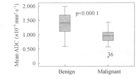
Fig.1 Comparison between ADC values of benign and malignant breast lesions.
ROC analysis was performed to achieve a feasible cut-off value of ADC for the differentiation of malignant and benign lesions.Breast lesions that had an ADC value at or less than the given cut-off value were considered malignant and lesions that had an ADC value more than the given cut-off value were considered benign.The value of area under the curve represents the probability that the lesion will be correctly classified as benign or malignant.ROC analysis revealed that the area under the curve was 0.861 and the cut-off value for ADC was 1.105 × 10⁻³mm2/s.At this value for ADC,the MRI protocol was found to have a sensitivity of 76%,a specificity of 78%,and a positive predictive value of 11.3% and a negative predictive value of 12.9%.
Kinetic analysis
On evaluation of the kinetic curves generated,it was found that 18.5%of the lesions with a type 1 kinetic curve were found to be malignant,81.8%of the lesions with a type II kinetic curve were malignant while 83.3%of the lesions with a type III kinetic curve were malignant.
4 Conclusion
In this study malignant lesions have significantly lower ADC values than benign lesions.The threshold ADC value between malignant and benign lesions is found to be 1.105 ×10⁻³mm2/s,with a sensitivity of 76%and specificity of 78%.Our study also demonstrates that type II and type III curves should be considered indicative of malignancy.
[1]Bartella L,Smith CS,Dershaw DD,etal.Imaging breast cancer[J].Radiol Clin North Am,2007,45(1):45-67.
[2]Kuhl CK.MRI of breast tumours[J].Eur Radiol,2000,10(1):46-58.
[3]Orel SG.High resolution MR imaging for the detection,diagnosis and staging of breast cancer[J].RadioGraphics,1998,18(4):903-912.
[4]Evis Sala,Suzanne Wakely,Emma Senior and David Lomas.MRI of malignant neoplasms of the uterine corpus and cervix[J].American Journal of Roentgenology AJR,2007,188(6):1577-1587.
[5]Heller D,Hricak H.Cost-effectiveness of new technologies for staging endometrial cancer[J].Eur Radiol,2000,10(3):381-385.
[6]Belli P,Costantini M,Bufi E etal.Diffusion-weighted imaging in breast lesion evaluation [J].Radiol Med,2010,115(1):51-69.
[7]Bogner W,Gruber S,Pinker K,etal.Diffusion-weighted MR for differentiation of breast lesions at 3.0 T:how does selection of diffusion protocols affect diagnosis?[J].Radiology,2009,253(2):341-351.
[8]Guo Y,Cai YQ,Cai ZL,etal.Differentiation of clinically benign and malignant breast lesions using diffusion-weighted imaging [J].J Magn Reson Imaging,2002,16(2):172-178.
[9]Kinoshita T,Yashiro N,Ihara N,etal.Diffusion-weighted half-Fourier single-shot turbo spin echo imaging in breast tumours:differentiation of invasive ductal carcinoma from fibroadenoma[J].J Comput Assist Tomogr,2002,26(6):1042-1046.
[10]Kuroki Y,Nasu K,Kuroki S,etal.Diffusion-weighted imaging of breast cancer with the sensitivity encoding technique:analysis of apparent diffusion coefficient value[J].Magn Reson Med Sci,2004,3(2):79-85.
[11]Marini C,Lacconi C,Giannelli M,etal.Quantitative diffusion MR imaging in the differential diagnosis of breast lesion [J].Eur Radiol,2007,17(10):2646-2655.
[12]Rubesova E,Grell AS,De Maertelaer V,etal.Quantitative diffusion imaging breast cancer:a clinical prospective study [J].J Magn Reson Imaging,2006,24(2):319-324.
[13]Sinha S,Lucas-Quesada FA,Sinha U,etal.In vivo diffusion weighted MRI of the breast:potential for lesion characterization[J].J Magn Reson Imaging,2002,15(6):693-704.
[14]Woodhams R,Matsunaga K,Iwabuchi K,etal.Diffusion-weighted imaging of malignant breast tumours:the usefulness of apparent diffusion coefficient(ADC)value and ADC map for detection of malignant breast tumours and evaluation of cancer extension[J].JComput Assist Tomogr,2005,29(5):644-649.
[15] Woodhams R,Matsunaga K,Kan S,etal.ADC mapping of benign and malignant breast tumours[J].Magn Reson Med SCI,2005,4(1):35-42.
[16]Patrick Z.McVeigh,Aejaz M.Syed,Michael Milosevic,Anthony Fyles,Masoom A.Haider Diffusionweighted MRIin cervical cancer[J].Eur Radiol,2008,18(5):1058-1064.
[17]Beatty JD,Porter BA.Contrast-enhanced breast magnetic resonance imaging:the surgical perspective[J].Am JSurg,2007,193(5):600-605.
[18]Esserman LJ,Kumar AS,Herrera AF,etal.Magnetic resonance imaging captures the biology of ductal carcinoma in situ[J].J Clin Oncol,2006,24(28):4603-4610.
[19]Kuhl C,Kuhn W,Braun M,Schild H.Pre-operative staging of breast cancer with breast MRI:one step forward,two steps back?[J].Breast,2007,16(2):34-44.
[20]Teifke A,Behr O,Schmidt M,etal,Dynamic MR imaging of breast lesions:correlation with microvessel distribution pattern and histologic characteristics of prognosis[J].Radiology,2006,239(2):351-360.
[21]Szabo BK,Aspelin P,Kristoffersen Wiberg M,Tot T,Bone B.Invasive breast cancer:correlation of dynamic MR features with prognostic factors [J].Eur Radiol,2003,13(11):2425-2435.
- 外科研究與新技術的其它文章
- Meta-analysis of the efficacy between gemcitabine-alone and gemcitabine-based chemotherapy applied to advanced pancreatic cancer
- Effect of decompression in different time on hemodynamics and oxygen metabolism of porcine model with severe acute pancreatitis combined intraabdominal hypertension
- Postoperative use of Sorafenib in liver transplantation patients of hepatocellular carcinoma beyond Milan criteria
- Combined liver-kidney transplantation and orthotopic liver transplantation in the treatment of severe hepatitis B
- HO-1 up-regulation decreases liver ischemia/reperfusion injury by inhibiting mast cells degranulation
- Laparoscopic cholecystectomy plus exploration of the common bile duct in comparison with laparoscopic cholecystectomy and endoscopic sphincterotomy for secondary choledocholithiasis